Forschung

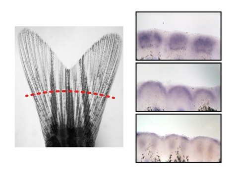
Zebrafish caudal fin (left). Gene expression in the early regenerating fin (right).
Although bone is a regenerative tissue also in humans, it can be permanently lost in circumstances of trauma, cancer or degenerative diseases. Due to their high regenerative capacity zebrafish have emerged as a powerful tool to study bone regeneration. In particular, bone forming and bone degrading cells (osteoblasts and osteoclasts) can be visualized easily in zebrafish, thus allowing their observation in the living organism. This can be used to monitor osteoblast and osteoclast function throughout processes of bone formation and bone degradation, and to study cross-talk of bone cells with other types of cells, such as immune cells, in the bone niche.
In the lab we make use of pharmacologic approaches and molecular biology tools to investigate adverse effects of certain drugs used in the clinics. As an example, we have analyzed the anti-regenerative effects of immunosuppressive glucocorticoids in zebrafish, in which bone forming osteoblasts are fluorescently labelled. In doing so, we found that both osteoblast activity and proliferation are reduced by glucocorticoid exposure, while osteoblast apoptosis remains unaffected. We now aim to identify molecules which counteract bone destruction in this context and hope that this will give novel impulses in the treatment of inflammatory diseases.
A main focus of the lab is to understand underlying mechanisms of succesful bone regeneration. Zebrafish regenerate bone quickly after injury, both in appendages and the skull; however, they do not overgrow bone. How do they do so? To address this question, we explore ways of positively and negatively influencing tissue regeneration. This is feasible both by drug treatments and genetic modulation. By using a combination of these approaches with state of the art imaging technologies, we hope to identify mechanisms that are involved in growth control of bone and of regenerating tissues in general. This may have important implications for future regenerative therapies targeting bone.
