3D scanning of chronic wounds and scars to assess the course of treatment
| Runtime | 01.09.2010 - 31.08.2012 |
| Funding | Bundesministerium für Wirtschaft und Technologie in the AiF-program „Zentrales Innovationsprogramm Mittelstand“ (ZIM). |
| Project staff | Dr.-Ing. Stefan Holtzhausen |
| Partner | Uniklinikum Dresden: Poliklinik für Dermatologie |
Objectives
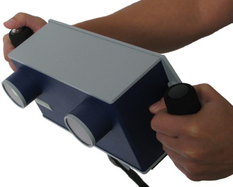
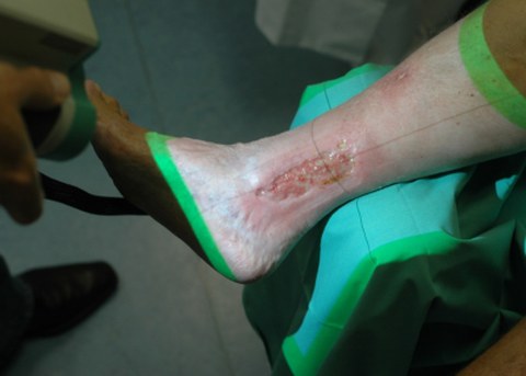
Special 3D scanners are used to detect skin defects. These work contact-free according to the fringe projection method and enable the scanning of surfaces in a very short period of time (t < 1sec) with sufficient resolution and accuracy. This makes it possible to record skin defects such as scars, ulcers, etc. directly on the patient. The scanners (e.g. Z-Snapper from Vialux) can be guided by hand and do not require complex laboratory conditions.
Project content
Hand-held 3D scanner zSnapper portable
(ViALUX Messtechnik + Bildverarbeitungs GmbH)
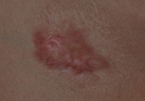
Proliferating scar
Patient with ulcers during admission with the Z-Snapper
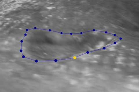
Textured 3D surface with interactively defined wound margin by the doctor
Determination of the wound or scar volume in wounds and scars. The healthy skin surface is approximated on the basis of the wound edge areas and the volume is determined by means of the digitised wound surface.
Results
Based on the recorded data, it is possible to determine the wound margin in the form of a 3D curve. This is then used to determine the surface area and to calculate the volume of the wound/scar. For this purpose, the surface shape of the healthy skin surface is estimated. Subsequently, the area of the scar/wound surface is compared with the estimated healthy skin surface.


