Histology
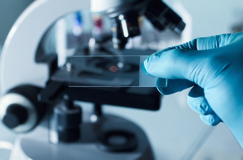
Our platform uses modern histological methods to image molecules and cells in tissue, thereby supporting scientists in the study of brain tumors, neurodevelopment and neurodegeneration. Various techniques such as immunohistochemistry, in situ hybridization and tissue clarification (light sheet microscopy) are used in the histology platform.
Immunohistochemistry is a method for visualizing specific proteins within tissues through binding with antibodies, while in situ hybridization allows for the detection of specific RNA or DNA sequences within tissues. Both techniques are crucial for studying molecular and cellular changes that occur physiologically as well as in brain tumors, during neural development and neurodegeneration.
Tissue clearing, on the other hand, is a relatively new technique that allows our scientists to make entire organs or tissues transparent to visualize their inner workings in three dimensions. Lightsheet microscopy is used to scan the cleared tissues at high resolution, allowing for detailed analysis of cells and structures. Together, these techniques offer unprecedented opportunities to understand the complex mechanisms underlying brain tumors and to analyze the normal processes of brain development and aging. The histology platform is therefore an indispensable resource for our scientists studying the brain and its many mysteries.

