C.elegans meiosis
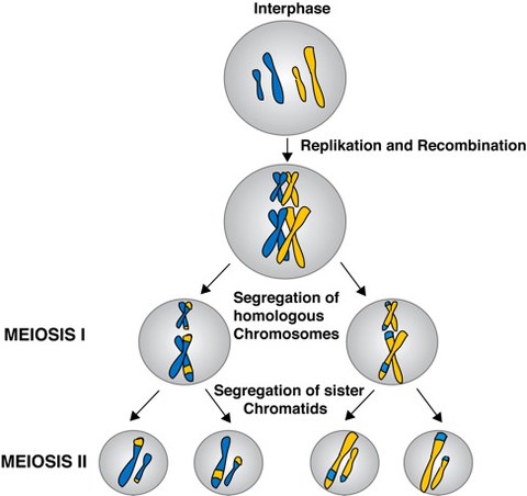
During fertilization, an egg and a sperm fuse to form a new embryo. Prior to this process the egg has to undergo a maturation process called meiosis (Figure 1). During meiosis, haploid gametes are produced from a diploid progenitor cell. This process occurs in all sexually reproducing eukaryotes. This reduction of the chromosome number is essential for the further development of the fertilized oocyte into an embryo. Defects in meiosis result in aneuploidies, leading to embryonic lethality and genetic disorders such as Down´s syndrom.
The machinery used for chromosome segregation in meiosis and mitosis relies on microtubules, motor proteins and other microtubule associated proteins. In contrast to mitotic spindles and meiotic spindles in male germ cells, in which microtubules are often organized by centrosomes, female meiotic spindles of many species, including C. elegans, Drosophila, Xenopus, mice, humans are anastral and acentrosomal. Female meiotic spindles must assemble and segregate chromosomes without microtubule organizing centres. This gives rise to several questions. How are the microtubules nucleated without centrosomes, and how do they manage to arrange into two poles? How are chromosomes segregated in the absence of spindle poles. By today the formation of meiotic spindles and the process of chromsome segregation is not very well understood. A major reason is the small size of the meiotic spindle, impeding the analysis of the structure by light microscopy.
We want to investigate the mechanisms of meiotic spindle assembly and chromosome segregation in the nematode C. elegans as a model organism.
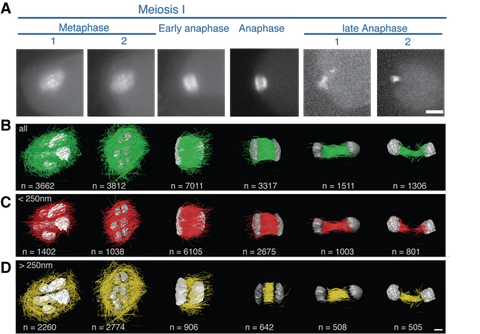
At an early metaphase stage in female meiosis in C. elegans, microtubules form an elongated bipolar spindle. During metaphase the spindle shortens and adopts a barrel-like shape. Once anaphase begins the microtubules mainly localize to the zone between the separating chromatids (Figure 2). The mechanisms of this microtubule reorganization during the metaphase-to-anaphase transition, as well as the detailed mechanism of chromosome segregation, however, are poorly understood.
For this we are using a combination of light microscopy and serial-section electron tomography to analyze the dynamics and ultra structure of the meiotic spindle in meiosis I and II (Figure 3).

Figure 1: Schematic overview of Meiosis I and II
Figure 1: Schematic overview of Meiosis I and II
Overview of the different stages of meiosis I and II. Following interphase the DNA of cells is replicated and the homologous chromosomes are aligned, allowing the recombination of genetic material. During meiosis I the homologous chromosomes are segregated into daughter cells. Meiosis I is followed by meiosis II, in which the sister chromosomes are segregated into daughter cells, giving rise to haploid cells.

Figure 2: Overview meiosis in C. elegans
Figure 2: Overview meiosis in C. elegans
At an early metaphase stage in female meiosis in C. elegans, microtubules form an elongated bipolar spindle. During the course of metaphase this elongated spindle shortens, adapting a barrel-like shape. With the onset of anaphase microtubules mainly localize to the zone between the separating chromatids.

Figure 3: Reconstructions of female meiotic C. elegans spindles
Figure 3: Reconstructions of female meiotic C. elegans spindles
A, Light microscopic images of the different meiotic C. elegans embryo just prior high pressure freezing. Scale bar 10 µm B, complete reconstructions of different meiotic spindles, number of microtubules is indicated in the bottom left corner. C, selected microtubules of the different datasets, which are located 250nm and closer to the chromosomes. D, selected microtubules of the different datasets, which are located further than 250nm from the chromosomes. Scale bar 1µm.
