Dec 15, 2025
Publication "Depolarization-Based Multimodal Optical Imaging of Carious Lesions"
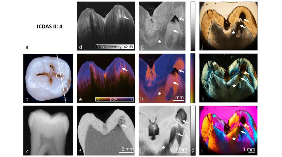
This publication presents a comprehensive, multimodal image catalog of occlusal caries lesions on teeth ex vivo, visually documenting different stages of caries progression.
The aim was to visualize caries using different optical imaging techniques in order to provide a reference basis for the development and validation of polarization-sensitive optical coherence tomography (PS-OCT) as a radiation-free diagnostic method. The study combines histological analyses, digital bitewing images, micro-CT and PS-OCT, with a particular focus on the evaluation of the degree of polarization (DOP).PS-OCT enables high-resolution, non-invasive cross-sectional images of hard dental tissue and uses the depolarization of light caused by demineralization in the enamel as a diagnostic contrast. The results show that DOP-based PS-OCT reliably detects initial caries lesions and can distinguish them from external discoloration or calculus. The method is particularly advantageous in the detection of non-cavitated, early caries sites, which are often not visible with conventional methods. In addition, the precise spatial registration of all image procedures in identical cross-sectional planes enables direct comparison and improves the interpretation of the data.
In summary, the publication provides a valuable, multimodal image catalog that serves as a reference for research and dental education. PS-OCT with DOP analysis shows great potential for the future, radiation-free diagnosis and monitoring of occlusal caries - provided that further clinical studies under real conditions follow.
https://onlinelibrary.wiley.com/doi/10.1002/jbio.202500422