Chromosome segregation in C. elegans male meiosis
Unlike the female meiotic spindle, the male meiotic spindle in the nematode Caenorhabditis elegans is assembled in the presence of centrosomes. Separation of homologous chromosomes in meiosis I and of sister chromatids in meiosis II gives rise to four equivalent haploid sperm cells, each containing DNA, a pair of centrioles, mitochondria and sperm-specific organelles. Applying both, live-cell imaging and electron tomography, we aim to characterize the dynamics and architecture of wild-type and mutant male meiotic spindles in C. elegans to understand how chromosome-segregation is achieved in primary and secondary spermatocytes.

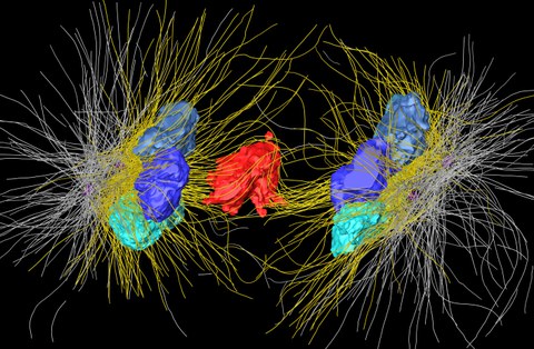
Three-dimensional reconstruction of a spindle in male meiosis 1 in males of the nematode C. elegans.
A key characteristic during male meiosis I is that a single unpaired sex chromosome is always segregated after the autosomes have been partitioned to the newly forming secondary spermatocytes. This offers a unique opportunity to probe segregation mechanisms on a single-chromosome level. We study this process applying genetic (mutants) and mechanical perturbations (laser microsurgery) and trying to obtain a detailed understanding by analyzing the ultrastructure of the involved cytoskeleton (microtubules, membranes, organelles) with electron tomography.
 © Stephan Wiegand
Postdoc
NameDr. Gunar Fabig
Send encrypted email via the SecureMail portal (for TUD external users only).
Visiting address:
Medizinisch Theoretisches Zentrum, Room: A.10.024 Fiedlerstraße 42
01307 Dresden
