Spindle organization in the trioecious nematode A. rhodensis
The nematode Auanema rhodensis (in older publication also found under the previous name Rhabditis sp. SB347) differs in many general characteristics from the well-studied model organism Caenorhabditis elegans: first, A. rhodensis is a trioecious nematode and shows three phenotypical sexes - males, females and hermaphrodites; second, it shows skewed sex ratios which do not follow Mendel´s laws; and third, there is an unusual segregation pattern during meiosis in male worms.
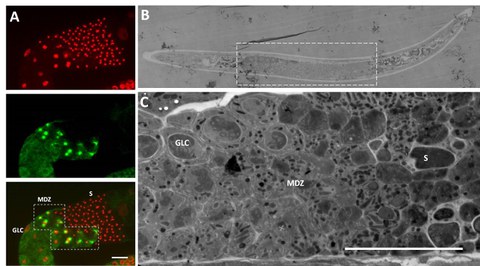
The examination of this uncommon pattern of the reductional division is part of my project. In order to analyze male meiosis within A. rhodensis males, different microscopic techniques are used (Figure 2). Light microscopy enables to get an overview about the anatomy of the males and to detect kinetochore as well as centriolar proteins through immunostainings. Electron microscopy of 70nm thin and 300nm semi-thick sections is used to obtain information about the ultrastructure of the meiotic cells and electron tomography of 300nm semi-thick sections allows to reconstruct different meiotic spindles in different stages of cell division. With the help of these reconstructed 3D models of meiotic cells we were able to analyze spindle structure, chromosome alignment and organelle distribution during male meiosis.
Particular attention was focus to anaphase II. In meiosis II, the X-chromatid lags and is distributed to just one of the two developing spermatids. Furthermore, an obvious asymmetric organelle distribution could be observed. The mechanism of this asymmetric distribution of both, the sex chromosome and the organelles is still not fully understood and the trigger of this redistribution remains to be elucidated.

Fig. 1: Different microscopic techniques are used to examine the male meiosis in A. rhodensis. (A) Immunostaining of α-tubulin (green) and labeling of DNA (red). The meiotic division zone (MDZ) is easily identifiable by the formed spindles. Proximally to this region germ line cells (GLC) are arranged while the zone of mature sperm (S) is distally located to the MDZ. (B) Bright field microscopy of a 300nm semi-thick section was used to find the meiotic region (dashed box). (C) The ultrastructure of meiotic dividing cells as seen in the MDZ, germline cells (GLC) and sperm (S) could be examined through electron microscopy. Scale: 10µm.
 © A. Schwarz
PhD student
NameAnna Unger
Send encrypted email via the SecureMail portal (for TUD external users only).
Visiting address:
Medizinisch Theoretisches Zentrum, A 10.024 Fiedlerstraße 42
01307 Dresden
