Spindle architecture in mono astral mammalian spindles
Faithful chromosome segregation relies on a precisely balanced network of forces within the mitotic spindle. In this project, we perturb these forces by inhibiting the outward sliding activity of the motor protein kinesin‑5 (Eg5). Loss of Eg5 function causes spindle collapse, resulting in a mono‑astral spindle in which the two poles fail to separate. By applying a small‑molecule inhibitor to block kinesin‑5 activity, we generate this controlled, tension‑reduced state, providing a powerful experimental system to analyze the organization of kinetochore microtubules (KMTs) and non‑KMTs in the absence of bipolarity.
Using quantitative analysis of serial‑section electron tomograms, we aim to uncover:
- What is the three‑dimensional (3D) organization of KMTs in Eg5‑inhibited spindles?
- How do non‑KMTs reorganize in mono‑astral spindles?
- How is the centrosomal architecture altered in collapsed spindles?
This work enables us to dissect the mechanics of spindle assembly by examining how the system reorganizes when a major force‑generating pathway is removed. Understanding these architectural principles will shed light on the fundamental mechanisms of KMT organization that ensure accurate chromosome segregation in mitotic cells.
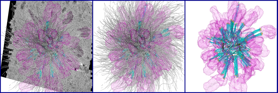
3D reconstruction and segmentation of a mono-astral spindle after serial-section electron tomography. Chromosomes shown in pink, KMTs in cyan, non-KMTs in gray, centrioles in green, KMT minus-ends in yellow, KMT plus-ends in red.
Contact
 © Stephan Wiegand
Postdoc
NameDr. Gunar Fabig
Send encrypted email via the SecureMail portal (for TUD external users only).
Visiting address:
Medizinisch Theoretisches Zentrum, Room: A.10.024 Fiedlerstraße 42
01307 Dresden