Structural analysis of abscission in C. elegans
Cytokinesis begins with signaling between the spindle and the cellular cortex. This signaling results in the generation of an equatorial zone, where a contractile acto-myosin ring assembles. As cytokinesis proceeds, an intercellular bridge with a densely packed center (called the midbody) and an overlap region of two bundles of antiparallel microtubules is formed. This region is called the intercellular bridge and the site of abscission, i.e. the final separation of the two post-mitotic daughter cells.
Current models of cytokinesis are mainly based on studies in cultured mammalian cells. Recently, we have visualized ESCRT-III-dependent filaments in HeLa cells in final stages of abscission (Guizetti et al., 2011). We could also show that abscission in mammalian cells in culture is accompanied by a disassembly of microtubules at the intercellular bridge.
Less, however, is known about cytokinesis in an embryonic system. We, therefore, want to investigate the mechanism of abscission in the early embryo of the nematode C. elegans. Using this model system, we could show that the intercellular bridge in the one-cell embryo C. is severed on both sides of the midbody (König et al., 2017). Thin spirals, as observed in mammalian cells, could not be detected. Using correlative light microscopy and serial-section electron tomography we will continue to analyze the dynamics and ultrastructure of this process in the early C. elegans embryo. In addition, we will study complete and incomplete events of cytokinesis in the gonad of this nematode species.
This is a collaborative project with the lab of Dr. Ana Xavier de Carvalho (Porto, Portugal).
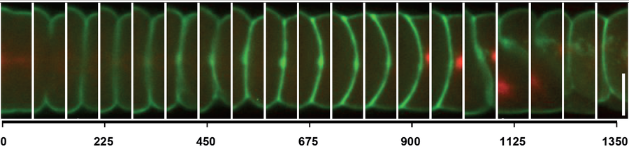
Figure 1: Fluorescence light microscopy of cytokinesis in the early C. elegans embryo. Scale bar, 10 µm.
 © wikimedia.org
Doktorandin
NameNicole Osang
Send encrypted email via the SecureMail portal (for TUD external users only).
Visiting address:
Medizinisch Theoretisches Zentrum, Room: A.10.026 Fiedlerstraße 42
01307 Dresden
References
König, J., Frankel, E. B., Audhya, A., and Müller-Reichert, T. (2017).
Membrane remodeling during embryonic abscission in Caenorhabditis elegans.
J. Cell Biol. 216, 1277–1286.
Guizetti, J., Schermelleh, L., Mäntler, J., Maar, S., Poser, I., Leonhardt, H., Müller-Reichert, T., and Gerlich, D.W. (2011).
Cortical constriction during abscission involves helices of ESCRT-III-dependent filaments.
Science, 331, 1616-1620.