Dec 17, 2024
EU millions for Dresden to develop portable quantum sensors: State of the art from quantum computing is set to open up new possibilities in neurosurgery

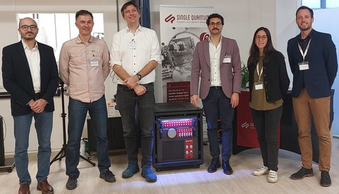
Prof. Tareq Juratli, Dr. Andriy Chmyrov, Prof. Oliver Bruns and partner (from left to right).
Optical measurement techniques are increasingly used in surgery for real-time tissue analysis. This technology is particularly important for monitoring during tumor removal, as it helps to detect any malignant cells that may still be present. These innovative techniques will also improve therapies for patients in the future. To this end, researchers from Dresden are collaborating with the Dutch company Single Quantum, which specializes in quantum technology, and Absolut System SAS, a French cryogenic engineering consultancy. They want to bring sensors that have so far only been used in quantum computing and communication to the operating room. An EU grant totaling EUR 5 million over four years is supporting the consortium in developing and implementing this technology.
The research alliance's joint project with the companies Single Quantum and Absolute System may sound a little like science fiction: Their aim is to harness a quantum sensor made of superconducting nanowire single photon detectors (SNSPDs) that is integrated into a time-resolved fluorescence microscope to improve the precision and completeness of neurosurgery for cancer operations. Put briefly, the incredibly sensitive sensor technology that is currently the norm in quantum information technology will be repurposed for use in operating theaters. The goal is to remove as much of the tumor as possible while sparing the surrounding tissue.
On the German side, three proven experts in imaging and neurosurgery are leading the PoQus research project. These are: Prof. Oliver Bruns, biochemist and head of the Department of Functional Imaging at the National Center for Tumor Diseases (NCT/UCC) Dresden and the German Cancer Research Center (DKFZ, Dresden site), Dr. Andriy Chmyrov, applied mathematician at NCT/UCC Dresden, and Prof. Tareq Juratli, Deputy Director of the Clinic and Polyclinic for Neurosurgery at the University Hospital Dresden (UKD) and Head of the Neurooncology Center at NCT/UCC.
“The use of this new technology can open up completely new prospects for our research in imaging and in neurosurgery,” Oliver Bruns and Tareq Juratli are convinced. “Integrating quantum sensors in neurosurgery promises to revolutionize tumor operations through highly precise, real-time imaging,” says Bruns. Juratli adds: “This technology enables more reliable detection of malignant cells while sparing healthy tissue, which has direct benefits for patient care.” The close collaboration between science and clinical application at NCT/UCC Dresden and UKD is a major asset in their eyes.
With specialized companies at their side, they have partners with extensive expertise in both quantum technology and cryotechnology for superconducting systems. Together, the consortium is ideally positioned to prepare and launch novel products on the market.
The technology
Real-time imaging must become more accurate. At the same time, it needs to be capable of penetrating deeper tissue layers. One solution is to combine two existing methods. Firstly, fluorescence lifetime imaging microscopy, which provides insights into the microenvironment of tumors and is therefore being used more frequently in cancer research. It allows tumor tissue to be distinguished from healthy tissue by exploiting the lifespan of fluorescent molecules in their excited states. Secondly, superconducting nanowire single photon detectors (SNSPDs) for higher time resolution and greater penetration depth. However, SNSPDs require intense cooling. The systems used so far are so bulky that they are unsuitable for clinical use.
“Using the SNSDPs as a basis, we want to develop a portable quantum sensor that is integrated into a time-resolved fluorescence microscope for use in operating rooms. The prototype will consist of a compact cryogenic cooling system, optimized detectors for maximum efficiency and speed, and software for image analysis,” says Oliver Bruns, outlining the plan.
The funding
The EU provides funding for research and innovation, particularly through the Horizon Europe program. PoQus is part of a funding line that only supports collaborative projects known as ‘Global Challenges and European Industrial Competitiveness,’ in Cluster 4: Digital, Industry and Space. PoQus will receive a total of EUR 4,986,529 in funding, with almost EUR 1.1 million going to the Faculty of Medicine at TU Dresden and around EUR 1.2 million to the German Cancer Research Center (DKFZ). This money also benefits the Dresden site through the DKFZ's financing of Oliver Brun's Chair at TUD's Faculty of Medicine.
About the researchers:
Oliver Bruns studied biochemistry and molecular biology at the University of Hamburg, which is also where he completed his doctorate. After working at the Leibniz Institute of Virology in Hamburg and the Massachusetts Institute of Technology (MIT, USA), Oliver Bruns came to the Helmholtz Pioneer Campus in Munich as Emmy-Noether-Group Leader of 'Next-Generation in vivo Imaging.' Since 2022, he has held a Chair at TU Dresden's Faculty of Medicine, which is funded by the German Cancer Research Center (DKFZ). He also heads the Department of Functional Imaging in Surgical Oncology at NCT/UCC Dresden.
Tareq Juratli completed his medical studies at Philipps-Universität Marburg, where he also received his doctorate. He began his residency at University Hospital Dresden (UKD) in 2007. Neurooncological research scholarships took him to Harvard University and Massachusetts General Hospital in the United States, before he returned to UKD in 2019 as a Senior Physician at the Department of Neurosurgery. Tareq Juratli heads the research group “Translational Neuro-Oncology and Skull Base Tumor Surgery” at the Department of Neurosurgery. He has been head of the Neurooncology Center at the NCT/UCC since 2022 and has served as Deputy Director of the Department of Neurosurgery since 2023.
Andriy Chmyrov studied applied mathematics at Sumy State University in Ukraine. He received his doctorate in physics from the KTH Royal Institute of Technology in Stockholm, Sweden, then conducted postdoctoral research at the Max Planck Institute for Biophysical Chemistry in Göttingen, Germany. Andriy Chmyrov has been working at Helmholtz Munich since 2015 – initially as a Group Leader at the Institute of Biological and Medical Imaging and, from 2021, as Head of the SWIR Microscopy Team. Since 2023, he has been Deputy Head of the Department of Functional Imaging in Surgical Oncology at NCT/UCC Dresden.
Contact:
Anne-Stephanie Vetter
Staff Unit Public Relations of the Carl Gustav Carus Faculty of Medicine of TUD Dresden University of Technology
National Center for Tumor Diseases (NCT/UCC) Dresden
+49 (0) 351 458 17903
