Label-free imaging of biological tissues
Vibrational spectroscopy is a method of optical analysis for the investigation of molecular structures and comprises infrared and Raman spectroscopy. Both methods are powerful analytical tools that can be used in a wide range of applications, from material characterization to medical diagnostics.
Our research focuses on the application of vibrational spectroscopy in oncology for the differentiation and characterization of tumor tissue. The advantages of infrared and Raman spectroscopy are that they are able to detect changes in cells and tissues at the molecular level without the need for all markers and staining. In addition, they are non-invasive and can therefore be carried out without damaging the tissue.
Raman spectroscopy has found an important application in neurosurgery to distinguish brain tumors from healthy brain tissue. In this area, we work closely with the Department of Neurosurgery and the Uckermann research group. The aim of our joint research is to develop new procedures so that the neurosurgeon can determine the position of the tumor more precisely during the operation in order to remove all of the affected tissue while sparing the healthy brain tissue as much as possible.
As part of the collaboration with neurosurgery, we have also developed a combined system for confocal Raman and Brillouin spectroscopy. Brillouin microscopy is an optical technique that enables the investigation of mechanical properties of materials at the microscopic level. We use it to study the biomechanics of cancer by non-invasively analyzing the viscoelastic properties of cells and the extracellular matrix. In addition, the combination of Brillouin and Raman spectroscopy offers the unique opportunity to correlate the biomechanical and biochemical properties to gain new insights into cancer progression and metastasis processes.
For a deeper insight into these research topics:
Galli et al. Rapid Label-Free Analysis of Brain Tumor Biopsies by Near Infrared Raman and Fluorescence Spectroscopy - A Study of 209 Patients. Front Oncol 9:1165 (2019).
Rix et. al. Correlation of biomechanics and cancer cell phenotype by combined Brillouin and Raman spectroscopy of U87-MG glioblastoma cells. J R Soc Interface 19(192):20220209 (2022).
Multiphoton microscopy offers innovative possibilities for multimodal visualization of anatomical and biochemical tissue information. It uses strong, focused laser beams to create nonlinear optical effects based on the interaction of multiple photons in a molecule.
Coherent anti-Stokes Raman scattering (CARS) microscopy is a nonlinear variant of Raman scattering that enables imaging based on the contrast created by the varying lipid content of the tissue. It offers chemical selectivity at subcellular resolution and high imaging speed. In addition, CARS imaging can be combined with other non-linear imaging techniques in the same system.
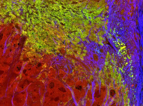
In our lab, we use a scanning microscope in combination with picosecond fiber lasers, which is suitable for different ex vivo or in vivo applications. The setup enables the simultaneous recording of CARS, two-photon fluorescence (TPEF) and frequency doubling (SHG). By superimposing the three levels of information, we obtain a more precise image of cells and tissues.
In collaboration with various clinics and research laboratories, we use multimodal CARS microscopy in oncology, cardiology and regeneration research as well as in basic biomedical research.
For a deeper insight into these research topics:
Galli et al. Identification of distinctive features in human intracranial tumors by label-free nonlinear multimodal microscopy. J Biophotonics. 12(10):e201800465 (2019)
Galli et al. Label-free multiphoton microscopy enables histopathological assessment of colorectal liver metastases and supports automated classification of neoplastic tissue. Sci Rep. 13(1):4274 (2023)
Galli et al. Label-free multiphoton microscopy reveals relevant tissue changes induced by alginate hydrogel implantation in rat spinal cord injury. Sci Rep. 8(1):10841 (2018).

Multiphotonenmikroskopie einer Probe von kolorektaler Lebermetastase. CARS (rot) wird für die Abbildung von Lipiden verwendet, TPEF (grün) für die Darstellung von zellulären Strukturen und SHG (blau) für die Visualisierung von Kollagenfasern. Durch das Zusammenführen der Informationen aus allen drei Kanälen entsteht ein umfassenderes Bild des Gewebes, welches zur Analyse der Mikromorphologie von Tumoren sowie zur präzisen Lokalisierung der Tumorgrenzen genutzt werden kann.
