Equipment
This is the equipment our group is currently working with. Please contact us for more information on our devices, collaboration or usage requests.
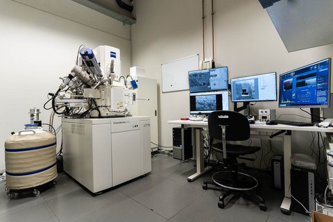
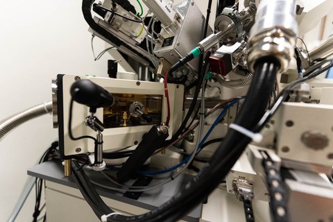
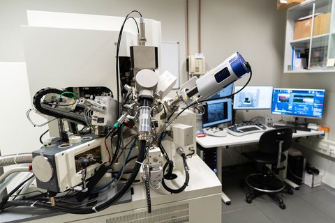
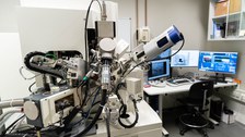
Cryo-Focused Ion Beam-Scanning Electron Microscope (FIB/SEM)
Crossbeam 550, Zeiss
Cryo-FIB/SEM combines imaging and nanofabrication technologies to perform volume imaging, material processing and sample preparation at the nanometric scale at room temperature as well as in cryogenic conditions.
Specifications - Electron Beam:
| Emitter: | Schottky Emitter |
| Beam current: | 1 pA – 100 nA |
| Resolution: | 1.8 nm @ 1 kV
0.9 nm @ 15 kV 0.7 nm @ 30 kV (STEM mode) |
Specifications - Focused Ion Beam
| Beam current: | 10 pA – 300 nA |
| Lifetime: | 3000 μAh |
| Resolution: | Other specs |
Other Specs
| Detectors: | Inlens, EsB, SESI, STEM, BSD |
| Stage: | X = 100 mm, Y = 100 mm, Z = 50 mm, T = -4° to 70°, R = 360° |
| Single GIS: | Pt, C |
| Scan field: | 32 k x 24 k (up to 50 k x 40 k with optional ATLAS 3D) |
| Micromanipulator: | Kleindiek Cryo-Gripper |
| Attachments: | Quorum PP3010T cryogenic preparation system (allowing to sputter coat the sample with a conductive layer in cryogenic conditions) & Plasma cleaner |
MPI-CBG, Room U47
Responsible Person: Luca Bertinetti
+49 351 463 44295
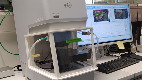
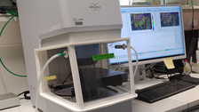
Bruker LUMOS II
| Detectors |
LN-MCT, smallest field of view 5x5 µm |
| Detector range |
4000 – 600 cm-1 |
| Operation modes |
Reflection, Transmission, ATR |
| Optics | ZnSe |
| Field of view | 1490 x 1118 µm2 |
B CUBE, Room 308
Responsible Person: Alice Ludewig
+49 351 463 44291
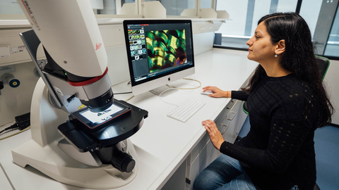
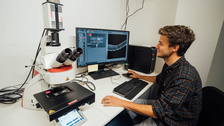
Leica DVM6, Digital Macroscope
| Objective: | PLANAPO with 12.55 FOV |
| Magnification: | 12x to 4740x, 16:1 zoom ratio |
| Camera: | 10-Mpixels high-resolution camera |
| Tilting: | From -60° to +60° |
B CUBE, Room 308
Responsible Person: Alice Ludewig
+49 351 463 44291
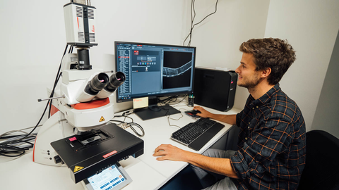
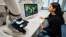
Leica DM6, Fluorescence Microscope
| Operation modes: | Bright field, reflection, Differential interference contrast, Polarized light and fluorescence |
| Objectives: | 1.25x, 5x, 10x, 20x, 40x and 63x |
| Filters: | 1 - GFP (Ex: 450 - 490 nm, DC: 495, Em: 500 - 550) 2 - 405 (Ex: 375 - 435 nm, DC: 445, Em: 450 - 490) 3 - TXR (Ex: 540 - 580 nm, DC: 585, Em: 592 - 668) |
B CUBE, Room 325
Responsible Person: Alice Ludewig
+49 351 463 44291
Linkam CMS196V³ Cryo-Correlative Microscopy Stage
| Operating temperature: | -196°C |
| Autonomy: | Up to a full day with 3L autofill LN2 Dewar |
| Stability: | No mechanical connection between sample mount and Dewar Low drift in nm range Good long-term stability |
| Stage: | Integrated motorised XY stage with position readout of better than 1µm |
| Optics: | Integrated condenser optics for transmitted light brightfield and phase contrast |
| Monitoring: |
Integrated temperature sensors |
MPI-CBG, Room U47
Responsible Person: Luca Bertinetti
+49 351 463 44295
ThermoGravimetric Analysis Differential Scanning Calorimetry (TGA/DSC)
Sensys Evo, Setaram
| Working T range: | Ambient to 830°C |
| Heating rate: | 0,01 to 30 °C min-1 |
| Sample volume: | 120 to 320 µl crucibles |
| RMS Noise: | 0,2 µW |
| Resolution: | 0,35 µW / 0,035 µW |
| Other features: | Adapted for water sorption isothermal measurements (with the Wetsys humidity generator) |
Room 308
Responsible Person: Gargi Joshi
+49 351 463 44297
Uniaxial Tensile tester
Portable for synchrotron in-situ application
| Custom built uniaxial features: | With in-house designed environmental chamber allowing for temperature and humidity control - room temperature to about 60°C - 5% to 90% relative humidity, depending on the temperature |
| Load Cells: | 5N, 50N |
B CUBE, Room 307
Responsible Person: Luca Bertinetti
+49 351 463 44295

