3 Germ Layer Differentiation
The 3 germ layer differentiation proves the pluripotency of an iPSC line by showing that it can generate cell types from all 3 germ layers: ectoderm, endoderm and mesoderm.
For ectoderm and mesoderm lineage, the customer can decide between two distinct differentiation assays:
Directed Monolayer Differentiation
Using the STEMdiff™ Trilineage Differentiation Kit, human iPSCs are differentiated in a monolayer to precursor cells of all 3 germ layers and stained with lineage-specific antibodies:
- Ectoderm: Neural transcription factor PAX6 and intermediate filament protein NESTIN are expressed in neural precursor cells after 6 days of differentiation.
- Endoderm: Transcription factor SOX17 is expressed after 4 days of differentiation.
- Mesoderm: Transcription factor BRACHYURY (T) is expressed after 4 days of differentiation.
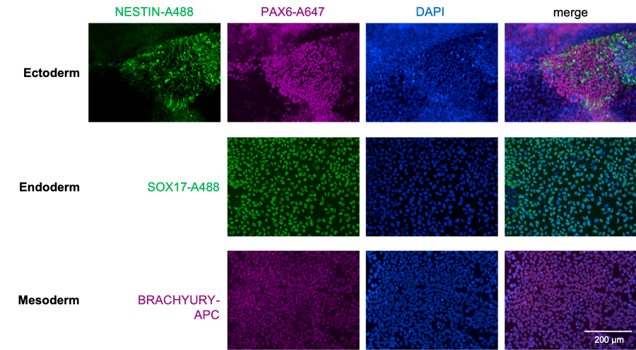
Human iPSCs differentiated in a monolayer to precursors of ectoderm, endoderm and mesoderm and stained with specific antibodies: PAX6, NESTIN, SOX17 or BRACHYURY (T). Scale bar 200 µm.
Spontaneous Differentiation via Embryoid Bodies (EBs)
Whole iPSCs colonies are detached from Matrigel using collagenase and cultured as floating aggregates for 8 days in medium containing FCS on non-adhesive dishes. After seeding of the resulting embryoid bodies (EBs) to gelatine, differentiating cells are growing out and are fixed on day 16 of differentiation. The inhomogenous mixture of different cell types is then stained for specific lineage markers:
- Ectoderm: Neuronal type 3 beta-Tubulin (TUJ1, TUBB3) identifies neurons.
- Mesoderm: Alpha-smooth muscle actin (SMA, ACTA2) stains vascular smooth-muscle cells.
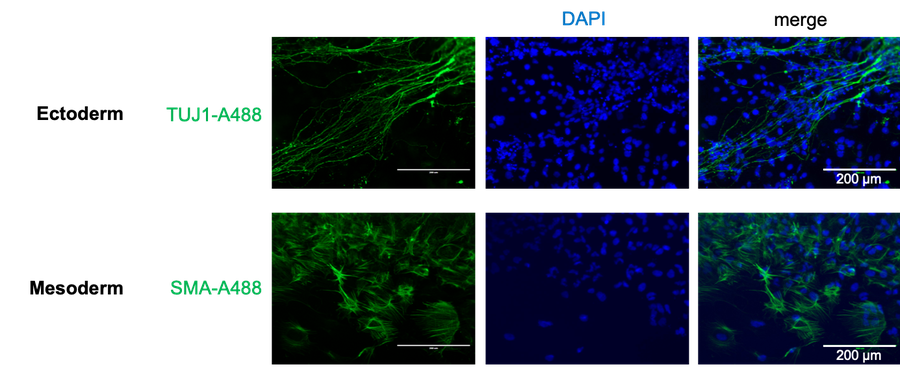
Human iPSCs differentiated for 16 days as embryoid bodies stained for the neuronal markers TUJ1 (TUBB3) and alpha-smooth muscle actin (SMA, ACTA2). Scale bars 200 µm.