Funded scientists 2023/2024
Table of contents
- Funded projects 2023/2024
- Dr. med. (Univ. Pécs) Felix Bechtolsheim
- Ani Cuberi
- Dr. rer. medic. Sarah Duin
- Dr. rer. medic. Sindy Giebe
- Dr. rer. medic. Robert Herber
- Dr. med. Moritz Herzog
- Dr. med. Julia Körholz
- Nora Martens
- Dr. med. Martin Mirus
- Dr. med. Lino Möhrmann
- Dr. med. Christoph Müller
- Dr. med. Leonora Pietzsch
- Dr. med. Sven Richter
- Dr. med. André Norbert Josef Sagerer
- Jonas Schadt
- Dr. med. Tom Alexander Schröder
- Dr. Alexander Schulz, Ph.D.
- Dr. med. Christian Teske
- Dr. rer. nat. Michele Wölk
- Dr. med. Christina Woopen
- Funded projects 2023/2024
Funded projects 2023/2024
Dr. med. (Univ. Pécs) Felix Bechtolsheim
Project title: Surgical Performance validated by Crowd-sourced Assessments of technical Skill “SuPer Crowd Skill”
Structural unit: Department of Visceral, Thoracic and Vascular Surgery
Project description
Oncologic surgery of the abdomen is associated with a high peri- and postoperative morbidity of patients. As recent research has shown, the intraoperative performance of the surgeon correlates significantly with the short term outcome (e.g. postoperative complications) as well as the long term outcome (e.g. overall survival) of the operated patients.
In this research project, the intraoperative performance of the surgeons will be measured by analyzing intraoperatively recorded surgical videos. The novelty of this project will be the usage of an AI-based motion analysis of the surgical instruments combined with a subjective assessment of surgical quality using validated scales (e.g. OSATS). As another novelty of this study, this subjective quality assessment shall be achieved by a so-called crowd-sourced assessments of technical skill (C-SATS). Here, "crowd" is understood as a large amount of raters without surgical expertise. Even with little guidance the sheer number of raters can achieve a high quality of assessment, comparable to the quality of surgical experts. The evaluation by the "crowd" will be performed online on anonymized video material, whereby such an evaluation by a sufficiently large "crowd" can be performed faster and in some cases better than a limited number of surgical experts could.
Ani Cuberi
Project title: Influence of thrombus composition and length on thrombectomy response- an in vitro study.
Structural unit: Institute and Polyclinic of Diagnostic and Interventional Neuroradiology
Project description:
Mechanical thrombectomy is the most effective method for treating proximal intracranial vessel occlusion in patients with ischemic stroke and has been the standard of care since 2015. Despite technical and procedural advances, complete reperfusion of the affected vascular territory cannot be achieved in approximately 20% of patients, which negatively affects the clinical course of patients. Several studies suggest that the composition, length and localization of the thrombus are associated with therapeutic response and the patients’ clinical course.
We aim to assess the influence of thrombus composition (fibrin-rich vs. erythrocyte-rich) and length on therapeutic outcome comparing different thrombectomy techniques and materials. Further, for different combinations of thrombus and thrombectomy technique, the volume of peripheral emboli – a common and devastating adverse effect during thrombectomy - will be recorded. Thus, novel insights for optimized thrombectomy will be obtained leading to a personal approach to interventional stroke therapy.
Dr. rer. medic. Sarah Duin
Project title: In vivo study of biocompatibility & functionality of neonatal porcine pancreatic islets in 3D bioprinted macroporous hydrogel constructs.
Structural unit: Centre for Translational Bone, Joint and Soft Tissue Research
Project description
While type 1 Diabetes mellitus can be adequately treated, it cannot really be cured and the vast majority of patients will develop long-term complications such as renal failure over the course of their lives, which so far can only be avoided by the transplantation of pancreatic islets from donors. To counteract donor shortage it would be advantageous to use islets from animals, ideally neonatal pigs. Due to large immunological differences between the species, in this case the islets necessarily have to be shielded from the human immune system which can be achieved by encapsulation in hydrogels, whereby a large surface decreases the diffusion distances nutrients, glucose and insulin have to cross and could possibly enable a quicker reaction. In the past, we were able to show than neonatal porcine islet-like clusters remain viable and functional when encapsulated in hydrogel-scaffolds prepared via 3D bioprinting.
Aim of the current project is now to transfer and expand this knowledge in an in vivo study to determine whether these constructs show immunoprotective capacities and whether the encapsulated islet-cluster can regulate blood glucose levels.
Dr. rer. medic. Sindy Giebe
Project title: Impact of maternal gestational diabetes and smoking during pregnancy on fetal vascular function
Structural unit: Department of internal Medicine III
Project description
Cardiovascular diseases are the leading cause of death worldwide and caused by a wide variety of risk factors. Smoking accounts for over 10% of cardiovascular-related deaths and is the most important avoidable risk factor for cardiovascular diseases.
In addition to direct effects on the vascular system, smoking also has a systemic effect in the body, impairs blood sugar levels, leads to insulin resistance and promotes the development of diabetes. The predisposition to cardiovascular disease in adulthood already starts before birth in the womb. Both, smoking during pregnancy and gestational diabetes (GDM), is associated with an increased risk of type 2 diabetes and cardiovascular complications later in life for mother and child.
The molecular and functional connections between smoking and GDM and their impact on vascular function and cardiovascular risk later in life have not been studied so far. In this project, placental vessels and isolated vascular endothelial cells will be analysed at molecular and functional level to contribute a better understanding of the effects of these risk factors on the cardiovascular system.
Dr. rer. medic. Robert Herber
Project title: The investigation of corneal biomechanics and hydration of corneal cross-linking by Brillouin microscopy
Structural unit: Department of Ophthalmology
Project description
The corneal cross-linking (CXL) is an established treatment procedure to stabilize the corneal tissue and halt the progression of keratoconus. Keratoconus is an ectatic disease of the cornea that is characterized by thinning and irregular bulging of the cornea leading to an irreversible loss of vision. During the CXL procedure, Riboflavin solution is applied to the corneal surface, which receives an UV light irradiation afterwards. The optimization of the Dresden protocol (standard protocol) is desired to provide the patients a more effective treatment. To investigate a potential effect of treatment modifications such as fluence, depth-dependent and quantitative information concerning the biomechanical enhancement of the tissue are necessary. The Brillouin microscopy is an optical measurement method that enables a depth-dependent quantification of biomechanical properties of the corneal tissue. The Brillouin shift is related to the longitudinal modulus (stiffness), which on the other hand depends from the hydration state of the tissue. The aim of this project is the investigation of the corneal hydration on the Brillouin shift and corneal stiffness to derive conditions that can be used in vivo as well. Pairs of porcine eyes will be used as specimens to perform CXL. Both eyes are treated with riboflavin, whereas only one will receive UV irradiation (complete CXL procedure). Afterwards, both eyes will soak in balanced salt solution to obtain the stable hydration. Brillouin and Raman spectroscopy will be performed to evaluate the process induced by CXL. Thereafter, a variation in corneal hydration will be achieved by different solutions. In the end, the investigations will be repeated in physiological conditions.
Dr. med. Moritz Herzog
Project title: Ultrasound spectroscopy: structured analysis of biomechanical tissue units
Structural unit: Department of internal Medicine I & Else Kröner Fresenius Center for Digital Health
Project description
Ultrasonic spectroscopy is the analysis of the vibration behavior of tissues at different frequencies. Compared to optical spectroscopy, it offers a significantly greater depth of penetration. The biomechanical properties obtained in this way have increasingly become the focus of research in recent years, as they could open up additional options for cancer diagnostics, for example. Isolated studies have already demonstrated the feasibility of ultrasound spectroscopy for ex-vivo and in-vivo scenarios. However, there is no systematic analysis of the frequency response of biological tissues. Rather, the studies show differences in individual sections of the frequency band, depending on the ultrasound equipment currently available. Using broadband MEMS technologies, this limitation can be overcome in the laboratory to establish a systematic study with significantly larger spectra. With the help of clinically approved ultrasound devices, which have a raw data interface, a transfer of this knowledge into clinical application should be achieved.
Dr. med. Julia Körholz
Project title: Delayed thymic maturation as a consequence of fetal growth restriction
Structural unit: Department of Paediatrics
Project description
Children with fetal growth restriction (FGR) are more frequently affected by susceptibility to infections or chronic diseases. This observation can be partly attributed to reduced thymic maturation caused by undersupply during pregnancy. Catch-up growth probably does not occur to a sufficient extent in this situation. T-cell development takes place in the thymus. Impaired T-cell maturation may lead to gaps in the host defense on the one hand and to overactivation of the immune system on the other hand. Longitudinal examination of specific T cell populations and sonographic monitoring of thymus size will be used to investigate impaired thymic development. Clinical data will be correlated to immunological data and FGR infants will be compared to healthy newborns. Thus, the exact pathogenesis of impaired thymic maturation in children with fetal growth retardation will be better understood.
Nora Martens
Project title: XYZ monitoring: orthogonal ECG derivation in the monitoring of critically ill patients
Structural unit: Department of internal Medicine I & Else Kröner Fresenius Center for Digital Health
Project description
The ECG as a central sensor for monitoring patients is ubiquitous in the clinic. Up to now, a distinction has been made between a diagnostic ECG and an ECG for monitoring critically ill patients. Up to 17 electrodes are used for diagnostic purposes. For monitoring, this is too laborious and error-prone and 5 electrodes are used. The interdisciplinary research group PatientenFunk from medicine and electrical engineering has developed a wireless ECG prototype that derives orthogonally with 4 electrodes and by transformation shows 12 or more channels for a fully comprehensive diagnostics. This transformation has been known since the 1950s, but only modern computing power allows real-time transformation between the leads.
In the funded project, a transformation will be developed based on modern algorithms from machine learning. This enables real-time transformation and improves the validity of the derived ECG. In addition, a pathology analysis, especially myocardial infarction diagnostics, will be established on the vector level of the orthogonal leads. This allows a fully comprehensive diagnosis without the loss due to the selective display of specific leads. This technology allows continuous analysis of all ECG channels for monitoring critically ill patients while improving patient care with only 4 electrodes necessary.
Dr. med. Martin Mirus
Project title: Cardiac Arrest after LAE: vv-ECMO as an extension of the therapeutic options & establishment of a tailored-approach in the management of bleeding complications
Structural unit: Department of Anesthesiology and Intensive Care Medicine
Project description
The state-of-the-art therapy for fulminant pulmonary artery embolism (PE) is prolonged resuscitation with systemic lysis therapy and, if expertise is available, the establishment of extracorporeal cardiopulmonary resuscitation (eCPR) using veno-arterial ECMO. At the same time, the effects of systemic lysis therapy on the homeostasis of the patient's coagulation system are fulminant and persistent. This often leads to manifest and severe bleeding complications of the lysed patients, especially after arterial vascular punctures in the context of va-ECMO.
The aims of this project are: (1) to demonstrate a potentially lower-risk therapeutic option for fulminant PE with vv-ECMO and (2) to establish a point-of-care-guided therapy of bleeding complications of lysis therapy by means of thrombelastography in order to fundamentally improve both acute treatment and complication management.
Dr. med. Lino Möhrmann
Project title: Comprehensive molecular analysis of rare cancers
Structural Unit: National Center for Tumor Diseases (NCT/UCC) Dresden
Project description
Rare cancers are often difficult to treat; in particular, molecular pathogenesis-oriented medical therapies are lacking. As previously shown in cancer of unknown primary (CUP), comprehensive molecular characterization using whole-genome/exome sequencing and transcriptome analysis enables molecularly informed treatments that can lead to additional treatment options in a substantial proportion of patients (Möhrmann et al., Nature Communications 2022 and Horak et al., Cancer Discovery 2021).
In this project, we would like to analyze further rare cancers to determine the value of comprehensive molecular analysis using data from the NCT/DKTK MASTER program. MASTER is a continuously recruiting, prospective observational study that addresses younger adults with advanced-stage cancer as well as patients with rare tumors. It offers a central platform for multidimensional characterization based on whole genome/exome sequencing and transcriptome analysis. Furthermore, it has recently been extended to methylome and proteome analysis. A dedicated molecular tumor board discusses the results and recommends molecularly informed therapies. Our findings may pave the way for future clinical trials.
Dr. med. Christoph Müller
Project title: Complications in endoscopic middle ear surgery - evaluation and reduction of thermal effects by optical systems.
Structural unit: Department of Otorhinolaryngology, Head and Neck Surgery
Project description
The development of powerful rigid endoscopes as well as passive and active endoscope holding systems in the last 2 decades has led to a growing number of indications for minimally invasive mono- and bimanual endoscopic ear surgery (EES). As the performance of optical systems is increasing, their intraoperative emission of radiation of different wavelength - e.g. thermal radiation in the long-wave infrared range or light in the visible spectral range - is also increasing. Increased energy input can cause damage to critical structures in the surgical site ‘middle ear‘(e.g. facial nerve, cochlea, middle ear mucosa). This translational project investigates in vitro and in vivo the influence of emitted radiation on critical structures of the middle ear. The target parameters for the assessment of intraoperative effects (temperature change, blood flow velocity as well as blood volume flow) are recorded and evaluated by means of camera-based multispectral analysis, thermography as well as by thermal sensors.
Dr. med. Leonora Pietzsch
Project title: Immune response in children with B-cell deficiencies after SARS-CoV-2 infection or vaccination
Structural Unit: Pediatric Immunology, Department of Pediatrics
Project description
Children and adults with congenital or acquired B-cell deficiencies have no or inadequate antibody responses after vaccination or wild-type infection. Lack of antigen-specific antibody production makes such patients vulnerable to bacterial and also viral infections. Despite immunoglobulin substitution, chronic or recurrent infections are not uncommon, and approximately 10% of these patients succumb to chronic lung disease and pulmonary failure.
In this project, we will collect clinical and laboratory parameters from a B-cell deficient cohort and healthy controls in 5 German centers longitudinally over a period of 12 months. Symptoms of proven SARS-CoV-2 infection as well as results of PCR, antigen testing, antibody titers (where meaningful) will be documented. In parallel, we will perform comprehensive immunophenotyping in order to capture the vaccination effect at the cellular level during the entire observation period. For detailed detection of SARS-CoV-2 reactive T cells, we will stimulate cells up to five days. Inflammatory signaling molecules (including interleukins, interferons, TNF-α) will be quantified from all samples by Cytometric Bead Array (BD Biosciences).
The results of this study should contribute to the understanding of the immune response to SARS-CoV2 infection and/or vaccination in B-cell deficiency and thus also lead to (so far missing) recommendations for this patient group.
Dr. med. Sven Richter
Project title: Realization of Raman spectroscopy in stereotactic biopsies
Structural Unit: Department of Neurosurgery
Project description: For patients with brain tumors, rapid and reliable diagnosis is essential to enable adequate therapy. Especially in areas of the brain that are difficult to access, stereotactic biopsy is used to obtain tissue. Despite conscientious planning and execution, about every 20th biopsy remains inconclusive in addition to time-consuming histological processing.
This MeDDrive project revolves around the clinical translation of optical methods to differentiate brain tumors. For this purpose, a fiber designed for stereotactic biopsies is coupled to a near-infrared laser, which excites the tissue intraoperatively and in vivo and measures the so-called “Raman Shift” with a spectrometer. The underlying phenomenon of Raman scattering is based on a slight energy difference between excitation light and scattered light due to molecular bond vibrations and is unique to the investigated tissue. Raman shift is therefore also referred to as a "biological fingerprint" which makes it suitable for differentiating a wide variety of tissues. In the long term, this project aims on shortening the time to diagnosis and minimizing the rate of inconclusive biopsies in order to improve patient care.
Dr. med. André Norbert Josef Sagerer
Project title: Characterization of the tumor microenvironment of melanoma brain metastases
Structural unit: Department of Neurosurgery
Project description
Metastases are the most common tumors of the central nervous system. Malignant melanoma show the highest prevalence of cerebral metastases. With the advancement of targeted and immunotherapy, better treatment outcomes can now be achieved. Unfortunately, patients receiving such therapies often develop resistance and experience recurrence or progress. Current data suggest that the tumor-specific microenvironment has a significant influence on the development of resistance. The aim of the present project is to immunologically and molecularly characterize the tumor microenvironment of melanoma brain metastases.
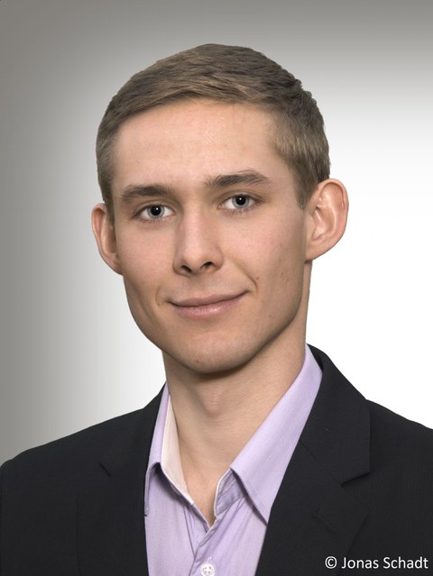
Jonas Schadt
Project title: Establishment of a flow cytometric method for the detection of measurable residual disease from peripheral blood in patients with acute myeloid leukaemia.
Structural unit: Department of internal Medicine I
Project description
Acute myeloid leukaemia (AML) still has a poor prognosis despite intensive therapies. The determination of measurable residual disease (MRD) below the light microscopic detection threshold by flow cytometry or genetic analyses from bone marrow aspirates is established for the early detection of relapse and for individual therapy management.
The aim of this project is to transfer the flow cytometric MRD-measurement algorithm for bone marrow to blood samples. This will not only enable less invasive diagnostics for the patient, but also the possibility of more frequent MRD monitoring. In this way, we hope to achieve better therapy individualisation and, as a result, a reduction of mortality.
Dr. med. Tom Alexander Schröder
Project title: Oxygenation and lactate real-time monitoring for enhanced tissue monitoring in free flaps: a novel electrochemical sensor prototype trial
Structural unit: Department of Oral and Maxillofacial Surgery
Project description
Flap surgery is a key element in any reconstructive surgical treatment, especially in maxillofacial cancer therapy. The oral cavity carcinoma is a major cause of morbidity and mortality in patients with head and neck tumor and furthermore one of the most common malignancies. To improve the functional outcome after surgical tumor removal, microvascular free flaps are the technique of choice.
The flap survival after surgery is paramount for the reconstruction of oral and facial tissue and of utmost importance to prevent facial disfigurement. In order to save the flap, in these cases, a quick re-exploration is necessary. Therefore, an early identification of flap failures has a top priority.
The requirement for any healing especially regarding free flaps is adequate tissue perfusion. The necessary nutrients and O2 can be monitored as well as their metabolic products. The main goal of the proposed study is to develop and validate a prototype of a monitoring sensor system which measures parameters like lactate and O2 saturation within the tissue. Through this approach, we will know what happens inside the flap and know its real physiology.
Dr. Alexander Schulz, Ph.D.
Project title: Understanding and overcoming therapy resistance of melanoma
Structural unit: Department of Dermatology
Project description
Melanoma represents the fifth most frequent malignancy in Germany and is the deadliest type of skin cancer. Activating BRAF mutations are detected in approximately 50% of all cases, thus representing the largest group by mutational status. For these patients, three different BRAF/MEK inhibitor combinations have been approved for treating to date, namely encorafenib+binimetinib, vemurafenib+cobimetinib, and dabrafenib+trametinib. However, the majority of patients develops resistance in less than two years. On the other hand, approximately 25% of melanoma cases harbor a mutation in the NRAS gene, being the second largest group of melanoma patients. In contrast to BRAF mutations, no efficient targeted therapies are available representing an urgent need for development of alternative strategies to improve survival of these patients. Resistance to existing therapies and lack of effective treatment remain a major obstacle in the treatment of many cancers, including melanoma. This research project aims to identify genes required for achieving and maintaining resistance in order to specifically inhibit their expression for (re)sensitizing these cells. Moreover, this project aims to screen clinically approved inhibitors for other diseases with tolerable safety profiles for anti-tumor activity against NRAS mutant melanoma, hopefully identifying a new treatment for this group of patients that can rapidly be transferred into the clinical setting.
Dr. med. Christian Teske
Project title: Biochemical profile analysis and differentiation of neoadjuvant chemotherapeutic pancreatic cancer organoids using molecular spectroscopic techniques.
Structural unit: Department of Visceral, Thoracic and Vascular Surgery
Project description
The ductal adenocarcinoma of the pancreas (PDAC) is one of the most lethal tumor entities among solid tumors. Recently, neoadjuvant treatment regimens before surgical resections are rising in the clinical setting in locally advanced PDACs. However, therapy response to chemotherapy is still heterogenous and not predictable. Human tumor organoids with molecular similarities to the primary tumor are currently evolving in biomedical and cancer research. The current project aims to analyze and classify the obtained PDAC organoids and their biochemical profile with spectroscopic methods, such as infrared spectroscopy, Raman spectroscopy and fiber-optical ATR-IR spectroscopy. The obtained data will be the basis for a chemotherapy response prediction and will lead to clinical correlation.
Dr. rer. nat. Michele Wölk
Project title: Dysregulated systemic release of metabolic and bioactive lipids along hepatocyte-VLDL axis
Structural unit: Center of membrane biochemistry and lipid research
Project description
Lipid mobilization, inter-organ transport and absorption within the body provide a functional readout of systemic lipid metabolism. The clinical lipid panel reports concertation of triglycerides, cholesterol, and phospholipids in different lipoprotein fractions (e.g., HDL, LDL, VLDL) to support diagnosis and prognosis of metabolic risks and disorders. Despite the clear significance of lipoprotein lipid component to systemic lipid metabolism, the lipoprotein lipidome and its remodelling in metabolic pathologies remain poorly characterized. Hepatocytes represent an excellent system to study the cross talk between cellular and systemic lipid metabolism, not only storing lipids in lipid droplets but also exporting lipids in form of verylow density lipoproteins (VLDLs). By using mass spectrometry based lipidomic approaches in a model system consisting of healthy and steatotic hepatocytes, this project aims to obtain a detailed molecular description of metabolic and bioactive lipids secreted in VLDLs under physiological and pathological conditions. This will enhance our current understanding on the liver – whole body metabolic cross talk in physiological and pathological conditions.
Dr. med. Christina Woopen
Project title: Characterization of Relapses in Multiple Sclerosis and Prediction of Therapeutic Potential: Cells, Hormones, Neurodestruction
Structural unit: Department of Neurology
Project description
Multiple sclerosis (MS) is a common chronic inflammatory disease of the central nervous system (CNS). MS is characterized by recurrent relapses in 85 % of patients, leading to neurological deficits that usually persist for several weeks before complete or incomplete remission. The pathophysiological basis is assumed to be the emergence of autoimmune dysfunctional immune cells and their invasion of the CNS. Therapies such as high-dose corticosteroids, plasmapheresis, immunoadsorption, or intravenous immunoglobulins can lead to an acceleration of symptom remission. In some patients, however, the therapeutic effect is insufficient. This project seeks to characterize MS relapses by means of a combination of innovative technologies with a focus on phenotyping of immune cells. Additionally, profiles of neuronal destruction markers and hormones during relapse and remission will be recorded. The aim of this study is the identification of a marker that is able to predict the response to relapse treatment.

