Nov 13, 2024
Novel finding on visual perception in autism: Differences in a peppercorn sized brain structure
Prof. Katharina von Kriegstein and her team at TU Dresden provide first direct evidence that autism is associated with different processing of visual stimuli in the magnocellular lateral geniculate nucleus (mLGN), a small but crucial structure in the brain. Their discovery was recently published in the prestigious journal Proceedings of the National Academy of Sciences (PNAS).
Autism is a neurodevelopmental condition that affects communication, interaction, and perception. Predominant theories on autism, however, often focus on communication and interaction. A recent study carried out by a team of neuroscientists at TU Dresden highlights the role of perception. For the first time, the researchers observed unique differences between autistic and non-autistic people in the magnocellular lateral geniculate nucleus (mLGN) The mLGN carries visual information from the eye to the cerebral cortex.
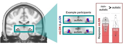
Using high-resolution functional magnetic resonance imaging (7T-fMRI), the team measured blood-oxygenation-level-dependent (BOLD) responses in the mLGN. This allowed to analyse the BOLD-responses and its differences between autistic and non-autistic adults in the mLGN. Results indicated reduced activity in autistic participants specifically in the mLGN, but not in other parts of the LGN.

The left side of the figure shows a section through a brain image with a map of the whole LGN (drop-shaped figures within the cyan rectangle). The cyan rectangle marks the insert that is shown in the middle of the figure and includes the mLGN (red) and pLGN (blue) from example participants. The pLGN is a part of the LGN which function was comparable for autism and control group participants. Right bar graph: Autistic participants had a lower response in the mLGN compared to the non-autistic control group.
One specialisation of the mLGN is the perception of motion. Motion also plays a role in social interaction and communication, such as in the perception of face movements when laughing or talking. Thus, differences in the mLGN could be a basis for parts of the characteristic social interaction and communication in autism.
“It is fascinating how fast and effortless our brain processes visual information. Much of this visual information is dynamic. For example, in face-to-face communication, the face movements of the communication partner can tell us something about what the person is saying or which emotional state the person has. The precise perception of these communication signals is an important part of social interaction,” explains Stefanie Schelinski, first author of the study. “The mLGN is tiny, comparable to the size of a peppercorn, and lies deep within the brain what makes it technically challenging to test this structure. In our study, we overcame technical challenges to test mLGN function and provide first direct evidence of its differencent functioning in autism. This opens up new perspectives for autism research. We hope that our findings will also inspire transdiagnostic research including clinical conditions that potentially overlap on a perceptual and neural level, such as schizophrenia and dyslexia.”
With a better understanding of the mLGN’s role, these findings could lay the groundwork for new diagnostics and improve the understanding of perceptual differences that are characteristic for autism.
Original publication
S. Schelinski, L. Kauffmann, A. Tabas, C. Müller-Axt, & K. von Kriegstein (2024). Functional alterations of the magnocellular subdivision of the visual sensory thalamus in autism, PNAS, 121(47), e2413409121. https://doi.org/10.1073/pnas.2413409121.
Contact:
Stefanie Schelinski
Research associate
Chair of Cognitive and Clinical Neuroscience
TU Dresden
E-mail:
Tel. (secretary Chair): +49 351 463-43901
Katharina von Kriegstein
Professor of Cognitive and Clinical Neuroscience
TU Dresden
E-mail:
Tel. (secretary Chair): +49 351 463-43901

