Jun 18, 2021
Interview with Dr. Marcus Jahnel – new research group leader at the BIOTEC and PoL

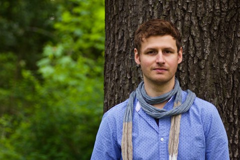
Dr. Marcus Jahnel
Dr. Marcus Jahnel started as a new research group leader at the Cluster of Excellence Physics of Life (PoL) and the Biotechnology Center of the TU Dresden (BIOTEC) on the 1st of June. His group “Dynamics of Biomolecules” uses the physics of light to study the physics of life.
How did your scientific journey look so far?
In 2006, I worked on my bachelor thesis on actin networks in the lab of Prof. Josef Käs at the University of Leipzig. Then, at the end of 2006, I moved to Manchester (UK) for a Master’s in Physics. I worked under Dr. Thomas Waigh on fibrin networks – networks of one of the main proteins involved in blood clotting.
In October 2007, I moved to Dresden to work on my PhD thesis “Single molecule studies of transcription” in Stephan Grill’s group at the Max Planck Institute of Molecular Cell Biology and Genetics (MPI-CBG). My work was supported by a grant from the Böhringer Ingelheim Fonds.
In 2013, I started my postdoctoral work and focused on three topics. With Loic Royer, who is now at the Chan Zuckerberg Biohub in the USA, I worked towards automating complex single-molecule experiments. In collaboration with Tony Hyman’s and Simon Alberti’s groups (MPI-CBG and BIOTEC), I studied the material properties of various biomolecular condensates. I also worked with Marino Zerial’s group and determined the function of long filamentous molecules, which can be found on specific cell organelles.
All these sound like biological projects. However, you are a trained physicist. What made you turn to Life Sciences and biophysics?
The interest, or rather a fascination, with biology was always there. But in school I also always enjoyed physics, its connection to mathematics, and the idea of being able to answer questions with clear, quantifiable answers. In retrospect, it might sound a bit naïve, but I liked that approach. Find a problem, describe it with equations, solve them, and you have a robust result that can be applied everywhere. Unfortunately, or fortunately, the reality is “a bit” more complex…
But then, biophysics sounds like a perfect solution! Do you remember what your first contact with biophysics was?
It was during my Bachelor’s thesis at the University of Leipzig. I remember seeing fluorescently labeled actin networks at the laboratory of Prof. Josef Käs. Actin is a polymeric protein that builds the cell’s skeleton. This was absolutely fascinating! Very different from the regular crystals, or the more abstract quantum phenomena that were otherwise in the physics curriculum. These experiments were easier to grasp and also easier to do ourselves. At the same time, we listened to lectures on polymer physics by Prof. Klaus Kroy, who described the behavior of polymers theoretically, with few elegant equations. So, I was moved by a mixture of exciting experiments that were visually very appealing, coupled with equations that described such complex behavior.
Both professors, Käs and Kroy, and their groups spread the mantra: physics and biology belong together! Physics can learn a lot from biology. What are the exciting questions? How does living matter differ from non-living matter? At the same time, physics offers a quantitative approach that tries to discover the truly essential properties of processes. Can we reduce something to the point where we can formulate it into an equation? On top of this, there are all those physical devices and intricate pieces of equipment (microscopes, lasers, atomic force microscopes, etc.) that made it possible to observe and measure things that had previously been hidden.
So investigating biologically motivated problems with a physical toolbox of experiments and theory has really drawn me in. It was only a logical step to go to a biophysics group for the Master's and the doctoral theses.
Now, 15 years later, you are starting your own biophysics research group to study biological condensates. How did you develop an interest in the condensates?
It was a coincidence. In summer 2008, I was sharing an office at the Max Planck Institute for the Physics of Complex Systems (MPI-PKS) with a postdoc from Tony Hyman's group. He just came back from a trip to a physiology course in the USA and was really excited: “Look at this! Isn’t this weird? It looks like honey drops, but it’s actually a protein!” He showed me the first videos of droplet-like protein aggregates that he made during the course. He was absolutely fascinated by this phenomenon. That postdoc was, of course, Cliff Brangwynne, who had just discovered together with Tony Hyman and Frank Jülicher the importance of phase separation of proteins that carry disordered regions. His excitement was contagious and I followed the development of the field with great interest from that point on. However, my research was still focused on transcription and single-molecule experiments.
A few years later, however, when the first phase-separating proteins were purified and became available in the laboratories of Tony Hyman and Simon Alberti, I became genuinely interested in working on them. The task now was to in-vitro characterize these novel states of matter of proteins. Do they behave more like water, like oil, or like honey? It was a wonderful coincidence that the protein droplets could be captured, moved, deformed, and measured with the optical trap – something I was very familiar with.
The experiments were relatively straightforward and, at the same time, yielded novel data that were very difficult to obtain by mere observation with a microscope. Droplets of all kinds are usually nice and round, so it is hard to tell from the pure image whether they are dynamic or more viscous. Controlled experiments with optical traps answer this very question and allow us to learn more about this state of matter. For example, with these experiments, we could show that the RNA-binding protein FUS – one of the proteins involved in neurodegenerative diseases like ALS – transitions from a fluid to a more viscous state and that this transition is accelerated in ALS mutants.
What do you find the most fascinating about the biological condensates?
One of the most exciting aspects is that they are dynamic and sometimes fragile assemblies that are largely determined by unfolded parts of a protein and weak interactions between them. It has long been taken for granted that the function of a protein is determined by its fold, i.e., its 3D structure. However, about 40% of human genes code for proteins with extended unstructured and unfolded regions. This somehow didn't quite fit into the bigger picture. Today, we understand that folded and unfolded protein regions are precisely balanced and that they have different levels of function. The dynamic collective behavior is often determined by the disordered parts, and then displays itself in phenomena such as phase separation and droplet formation. Many of these phenomena can be described quantitatively with thermodynamics and polymer physics.
You have mentioned an optical trap. It sounds like a special device. Can you explain how it works?
Sure! The research in my group is based on the ability to measure minute molecular forces, e.g., forces exerted by a single molecular motor as it runs, or forces needed to unfold a protein or an RNA, or forces required to stretch a small drop of condensate. We are talking about forces in the range of pN, i.e., forces that are only one-millionth of a millionth of the force a bar of chocolate exerts on a table it rests on (1 N). Mechanical instruments reach their limits and often cannot be reliably used here. Instead, we use light for our force measurements. One can measure such small forces with a focused beam of light and also use it to exert minute forces on a sample. The principle is called an optical trap or optical tweezers, and Arthur Ahskin was awarded a Nobel Prize in 2018 for its development. One can say that we use the physics of light to explore the physics of life.
Is the equipment already available on campus or do you have to set up the infrastructure for your group from scratch?
It’s already here. During my doctoral thesis in Stephan Grill's lab at the MPI-CBG, I developed, built, and programmed a high-resolution optical trap myself. This work took about three years and resulted in such a sensitive and robust measurement instrument that it is still used today.
After the PhD thesis, I supervised a PhD student who developed a second version of this optical trap at BIOTEC. This has the great advantage that it can be developed further and also automated. I will continue to use these two custom-built devices in my new group. Since these microscopes require a lot of know-how, there are not many researchers at the moment who can use them. This is definitely our advantage.
Are there also commercial setups available?
Yes! In the meantime, commercial optical traps have been developed that offer really high technical level. There are currently three such devices in Dresden – two at the MPI-CBG and one at the Molecular Imaging and Manipulation Facility at the Center for Molecular and Cellular Bioengineering. Again, having a background in performing single-molecule experiments and the associated data analysis is an advantage for an efficient use of these instruments. Therefore, I see our group as one of the main users and as natural collaborators for everyone interested in using the optical traps on the Dresden campus.
So it's fair to say that you're already well-positioned in terms of infrastructure. Is that correct?
Yes and no. One direction that I would like to establish soon, in addition to the optical traps, is microfluidics – for handling liquids and performing measurements on a tiny scale. There are many possible collaborations here with colleagues on campus and I am looking forward to diving deeper into this technology.
