Research fields
Our main research fields are optical coherence tomography, spectroscopy / multimodal imaging and thermography. Here you can learn more about these topics!
Optical coherence tomography (OCT)

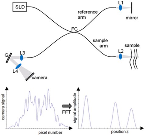
Schematic of a Fourier Domain OCT system.
The term „optical coherence tomography“ was first mentioned in the famous work of Huang and Fujimoto in 1991. OCT is a non-invasive and contactless method to image highly scattering subsurface samples up to a depth of about 1 mm with an axial resolution of 1 - 15 µm. Technically, OCT is based on low-coherence interferometry of the backscattered light from within the sample to measure the magnitude and echo time delay and with it depth information about the investigated object. Applications of OCT are biomedical research as well as clinical practice. Furthermore, more and more industrial applications are emerging, especially in non-destructive testing.
Our self-developed and built up OCT systems are all based on Fourier Domain OCT (FD OCT). First, the interference signal is spectrally analyzed and then the depth information is gained from the overall interference spectrum. We continuously enhance our OCT systems to improve especially the resolution and penetration depth into the medium of interest and to increase the image acquisition speed. In cooperation with other departments of the Faculty of Medicine and the University Clinic Carl Gustav Carus, different scanning units are developed and tested in clinical trials.
Spectroscopy and multimodal imaging

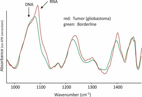
Infrared spectra of native, normal (tumor boundaries) and tumorous tissue. Significant differences in the range of the RNA bands can be seen
Optical molecular spectroscopy with their two most important representatives – Infrared- and Raman spectroscopy, is still the method with the highest information content of molecular structures. It is able to detect characteristics, which are based on structural properties of the sample, on a molecular level without additional markers. Thereby it fundamentally differs from microscopic test methods and labeling methods. While Raman spectroscopy has a high potential for in situ applications, Fourier Transform Infrared (FT-IR) Spectroscopy is mainly applied for ex vivo investigations of cells and tissues. By providing array detectors, the application potential has thereby considerably increased. By FT-IR imaging spectroscopy it is possible to determine the spatial distribution of chemical characteristics within one layer. In addition, methods of non-linear Rama spectroscopy allow fast imaging of tissues and cells. In combination with other optical imaging modalities, completely new insight into chemo morphologies and functions of biological material can be gained. A focus of our work is the histological characterization of tumors during surgical therapy.
The current focus concerning medical projects concentrates, among others, on the following main topics:
- Non-linear Raman spectroscopy imaging in neurosurgery and regenerative medicine
- In situ FT-IR spectroscopy in neurosurgery
- In situ Raman spectroscopy of tumor models in mice
- Spectroscopic differentiation of neural cells
- Examination of bone tissue
- Examination of collagen cross-linking in the cornea
- In ovo sex determination of chicken eggs

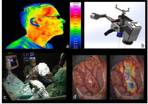
Thermography in neurosurgery. By using appropriate detectors and optics, thermal radiation of an object (wavelength range of 7 to 14 µm) can be made visible and its temporal change of heat distribution can be detected (a). Via a special adapter, the thermographic camera can be connected to a surgical microscope and can be used while neurosurgery such as tumor resection, epilepsy treatment or aneurysm treatment (b and c). Our aim is the intra-operational analysis of recorded thermograms to detect pathological changes in heat distribution of brain tissue and provide these information to the surgeon (d) during surgery.
Imaging techniques are of enormous importance in medical diagnostics. Thereby, the most interesting techniques are the ones that supply information on the non-visible spectral range (violet 380 nm up to red 780 nm wavelength), which cannot be detected by white-light cameras or the human eye. For example, MRT and CT show the inner structure of the body by utilizing the interaction of strong magnetic fields (MRT) or rather high-energy radiation (CT/x-ray) with biological tissue. Thermal radiation is a part of the non-visible spectral range (infrared radiation of 780 nm up to 50 µm wavelength), which is emitted by every object. This thermal radiation is not visible for common camera systems and the included information, like heat distribution and heat transport on the surface of an object, cannot be detected and used. By utilizing thermography systems with appropriate optical lenses and detectors, thermal radiation of an object and its temporal change can be made visible (e.g. thermal insulation of buildings).
There is an enormous potential for medical diagnostics, as biological tissue, depending on its cell structure and function, shows a different metabolism and thereby differentiated thermal changes. With high-sensitive cameras, differences in temperature of some millikelvin can be detected locally, whereby visualizing slightest difference of blood circulation and tissue activity is possible.
In cooperation with the Department of Neurosurgery of the University Clinic Dresden, we develop a method to make a localization of different types of tissue in the human brain possible by evaluating the temporal acquisition of heat distribution. As there are currently few possibilities to diagnose altered tissue structures discriminatingly, thermography is a promising method to enhance the patient care in neurosurgical interventions. In this project, we examine how the heat distribution of pathological tissue (e.g. tumor) can be distinguished from healthy tissue, how the blood flow of tissue sections and circulatory disturbances can be detected by thermography and how to possibly localize functional brain areas (sensory, motoric and associated areas).
