Oct 24, 2024
The great listener: Beta cell primary cilia and their various interactions
Similar to many other cell types in the body, pancreatic beta cells have a primary cilium, a protruding organelle that acts as a “cellular antenna.” Dysfunction of this organelle in beta cells has been linked to diabetes, however its structure and interactions have not been closely investigated. To address this, scientists at the Paul Langerhans Institute Dresden (PLID) of Helmholtz Munich at University Hospital and Faculty of Medicine of the Technische Universität Dresden, a partner of the German Center for Diabetes Research (DZD), the Human Technopole in Italy, and the Janelia Research Center and Yale University in the USA, have collaborated using various advanced imaging technologies to visualize the structure and interaction of beta cell primary cilia in their native environment. The results of this study, published in Nature Communications, give us a detailed look into the structure of these organelles and provide novel evidence of their connection to the nervous system.
Beta cells of the pancreas release insulin, the hormone required for glucose uptake from the bloodstream. Improper function of these cells leads to Type 2 Diabetes (T2D). There are many factors that could impair the ability of beta cells to produce insulin, making T2D difficult to combat. One factor that has recently been linked to T2D is the dysfunction of the primary cilia of beta cells. Most cell types in our body have a primary cilium, an organelle consisting of a protrusion of the plasma membrane with an underlying array of microtubules projecting from the cell body. Some types of cilia are motile and allow for cellular movement, while others, such as primary cilia, are usually non-motile but play important roles in extracellular signaling. Dr. Andreas Müller, in the Department of Molecular Diabetology (Director, Prof. Michele Solimena), led the team of researchers of various institutions who employed the use of volume electron microscopy (vEM), 3D segmentation, and ultrastructural expansion microscopy (U-ExM) in order to directly visualize the 3D form of beta cell primary cilia in their natural environment. The resulting images have provided new insights regarding the structure and function of beta cell primary cilia.
In this particular study, researchers addressed the question of how the axoneme, the skeleton-like structure formed by microtubules in the cilium, is organized. Depending on its function, the axoneme follows a specific pattern, either enabling the coordinated movement of the cilium or stabilizing the cilium without providing force for movement. The primary cilia observed in this study originated from both mouse and human beta cells and showed structural and molecular features of the latter case with dis-organized microtubules terminating at different distances within the cilium. These features could be observed in non-motile primary cilia from other cell types before, and could be shown for beta cells in this study for the first time.
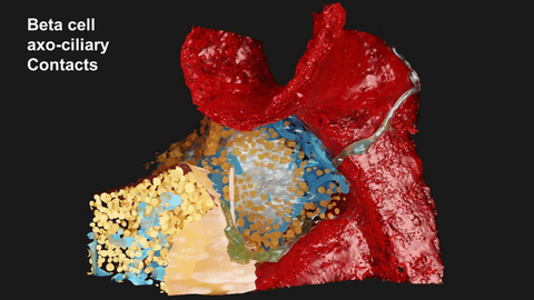
In addition to their inner structure, the authors were curious to find out how beta cell cilia interact with the cells surrounding them in order to make conclusions about their signaling capacity. First, they observed that beta cell cilia were spatially restricted by the surrounding tissue and made close contacts to neighboring cells. Due to this spatial restriction, primary cilia interact with adjacent cells as well as their cilia, sometimes contacting multiple cells at once. Further analyses of their imaging data indicated that beta cell primary cilia not only serve an important role in cell signaling and connectivity with other islet cells, but, surprisingly, with neurons as well. Islet nerves contacting beta cell cilia showed presynaptic characteristics indicating that primary cilia are also involved in neuronal signaling. Further testing showed that the majority of the connections between cilia and axons were positive for functional synaptic markers characteristic for acetylcholine signaling, an important neurological pathway for controlling islet function.
The structural data of this study reveals the importance of beta cell primary cilia as “major connective hubs pivotal for islet function” according to Müller. Further research into the mechanisms and pathways involved in primary cilia formation is now supported by the DZD Young Talent Program and will lead to a better understanding of their involvement in T2D pathogenesis.
Original publication:
Andreas Müller, Nikolai Klena, Song Pang, Leticia Elizabeth Galicia Garcia, Oleksandra Topcheva, Solange Aurrecoechea Duran, Davud Sulaymankhil, Monika Seliskar, Hassan Mziaut, Eyke Schöniger, Daniela Friedland, Nicole Kipke, Susanne Kretschmar, Carla Münster, Jürgen Weitz, Marius Distler, Thomas Kurth, Deborah Schmidt, Harald F Hess, C. Shan Xu, Gaia Pigino, Michele Solimena. Structure, interaction, and nervous connectivity of beta cell primary cilia. Nat Commun 15, 9168 (2024). https://doi.org/10.1038/s41467-024-53348-5

