Jun 20, 2023
A light in the darkness – illuminating the paths of protons

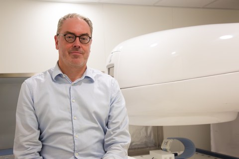
Prof. Hoffmann with the latest generation of in-beam MRI
The aim of proton beam therapy is to fight cancer by killing as many tumor cells as possible, while conserving the surrounding healthy tissue. As there is currently no way to see the beam during dose delivery, physicians work with safety margins around the tumor, which compromises the dose conformity and reduces target accuracy. Dresden researchers led by Prof. Aswin L. Hoffmann from OncoRay – the National Center for Radiation Research in Oncology have succeeded in visualizing the proton beam in a liquid-filled phantom using an in-beam MRI prototype and have developed this method to demonstrate the range of the proton beam during irradiation. The team published their results in the internationally renowned Journal Proceedings of the National Academy of Science. (DOI: 10.1073/pnas.2301160120)
Compared to photons, protons have a crucial advantage: they have a defined range, meaning a point at which they release their maximum energy. In proton radiation therapy, this attribute makes it possible to stop the radiation in the tumor tissue, and to apply a high dosage of radiation there, while greatly reducing the dose delivered to surrounding healthy tissue. For this reason, proton radiation therapy is mainly used to treat children as well as adults with tumors in the vicinity of tissue that is very sensitive to radiation. A method that directly measures and maps the range of the beam in relation to the patient's anatomy during dose delivery is essential for controlling dose delivery. As no such method has been used in routine clinical practice to date, safety margins have been built in around tumor tissue, resulting in the irradiation of healthy tissue and limiting the maximum possible dose to the tumor.
Prof. Hoffmann’s team has been researching the technical integration of magnetic resonance imaging (MRI) and proton therapy since 2016. Using an in-beam prototype, Hoffmann and his team have achieved a world first by visualizing the proton beam in a liquid-filled phantom and using this method to demonstrate the range of the proton beam during irradiation. “The results of our work could bring significant change to quality assurance in proton therapy. To this end, the proton beam is applied in water before the patient undergoes treatment, meaning that various beam properties can be checked in advance. Until now, these control measurements were indirect and time-consuming, but now proton beam imaging can happen directly during the measurement,” explains Hoffmann. “My dream is that this procedure will be used to monitor patient treatments in the future. To achieve this, we are looking for an MR procedure that displays the proton beam not only in water or other liquids, but also in human tissue.”
The feasibility study indicated that a visualization of proton beams in liquid is possible. As predicted, MRI images acquired during irradiation showed that the penetration depth increased with increasing proton energy, as did the MRI signal strength as the proton current increased. “This result is a significant leap in image-supported proton therapy,” said Prof. Mechthild Krause, Director of OncoRay. “Real-time MRI imaging has already made its way into conventional radiotherapy with photons. Professor Hoffmann and his team are working on the prototype of a new radiation device that will also establish real-time MRI imaging in proton therapy.”
The results of the group led by Hoffmann offer hope that a new dimension in the treatment of cancer patients will be made possible. A new large-scale MRI machine is currently being installed in the OncoRay building. This will make is possible for the first time to perform proton radiation and real-time MRI simultaneously, and also to vary the direction and strength of the magnetic field relative to the patient. This could allow proton therapy to be used even more precisely for moving tumors in just a few years, thus enabling the healthy tissue to be spared even more effectively and the tumor tissue to be irradiated with a higher dose.
Prof. Aswin Hoffmann
Head of Experimental MR-integrated Proton Therapy Research Group
at HZDR / OncoRay–
National Center for Radiation Research in Oncology
Stephan Wiegand
Public Relations & Marketing
Carl Gustav Carus Faculty of Medicine
TUD Dresden University of Technology
Tel.: +49 351 458-19389
Internet: http://tu-dresden.de/med/
