Feb 02, 2022
Using X-rays to Unveil the Secrets of Biofilms

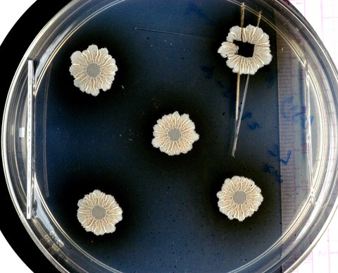
When bacteria join together to form communities, they may build complex structures. The photo shows wild-type Bacillus subtilis biofilms.
Most bacteria have the ability to form communities, biofilms, that adhere to a wide variety of surfaces and are difficult to remove. This can lead to major problems, for example in hospitals or in the food industry. Now, an international team led by Hebrew University, Jerusalem, and the B CUBE at the TU Dresden, has studied a model system for biofilms and found out what role the structures within the biofilm play in the distribution of nutrients and water.
Bacterial biofilms can thrive on almost all types of surfaces. They can be found on rocks and plants, on teeth, and mucous membranes, but also on contact lenses, medical implants or catheters, in the hoses of the dairy industry or drinking water pipes, where they can pose a serious threat to human health. Some biofilms are also useful, for example, in the production of cheese, where specific types of biofilms not only produce the many tiny holes, but also provide a delicious taste.
Tissue with special structures
"Biofilms are not just a collection of very many bacteria, but a tissue with special structures," explains Prof. Liraz Chai from the Hebrew University in Jerusalem. Together, the bacteria form a protective layer of carbohydrates and proteins, the so-called extracellular matrix. This matrix protects the bacteria from disinfectants, UV radiation, or desiccation and ensures that biofilms are really difficult to remove mechanically or eradicate chemically. However, the matrix is not a homogeneous sludge. "It's a bit like in a leaf of plants, there are specialized structures, for example, water channels residing in tiny wrinkles," says Chai. But what role do these structures play and what happens at the molecular level in a biofilm was not known until now. Prof. Chai joined forces with Prof. Yael Politi, research group leader at B CUBE – Center for Molecular Bioengineering at the TU Dresden, an expert in the characterization of biological materials, to apply X-ray diffraction, a non-destructive analysis technique available the synchrotron radiation source BESSY II at Helmholtz-Zentrum Berlin (HZB).
"The good thing about this technique and the instruments available at the HZB is that we could map quite large areas. By combining X-ray diffraction with fluorescence, not only could we analyze the molecular structures across the biofilm very precisely, but we could also simultaneously track the accumulation of certain metal ions that are transported in the biofilm and learn more about their biological roles," points out Prof. Yael Politi.
A model system for many biofilms
The scientists used biofilms from Bacillus subtilis, a harmless bacterium that thrives on plant roots and forms a useful symbiosis with them. It stores water so that the plant can possibly take moisture from the biofilm during drought and it also protects the roots from pathogens. In return, the cells in the biofilm feed on the liquids secreted by the roots. Bacillus subtilis bacteria can serve as a model system for many other bacterial biofilms.
At the MySpot beamline of BESSY II, the scientists examined a large area (in a range of square millimeters) of such biofilm samples. They were able to spatially resolve the structures within the biofilm and distinguish well between matrix components, bacterial cells, spores, and water. "X-ray fluorescence spectroscopy, is a method that allowed us to identify important metal ions such as calcium, zinc, manganese, and iron, even when present in trace amounts,” says Dr. Ivo Zizak, HZB physicist in charge of the MySpot beamline. This made it possible to correlate biofilm morphology and metal ion distribution.
Spore formation at unexpected locations
The results show that calcium ions preferentially accumulate in the matrix, while zinc, manganese, and iron ions accumulate along the wrinkles, where they can possibly trigger the formation of spores, which are important for the spread of the bacteria.
"We didn't expect that, because normally spores form under stress, e.g., when there is not enough water available. But here the formation of spores is actually linked with water channels, probably due to the accumulation of metal ions," says Chai.
The results show that the structures in the matrix not only play an important role in the distribution of nutrients and water, but also actively influence the bacteria's ability to behave as a multicellular organism. "This could help us to better deal with biofilms overall, with the beneficial ones as well as the harmful ones," says Liraz Chai.
Publication
David N. Azulay, Oliver Spaeker, Mnar Ghrayeb, Michaela Wilsch-Braeuninger, Ernesto Scoppola, Manfred Burghammer, Ivo Zizak, Luca Bertinetti, Yael Politi and Liraz Chai: Multiscale X-ray study of Bacillus subtilis biofilms reveals interlinked structural hierarchy and elemental heterogeneity. PNAS (January 2022)
Link: https://doi.org/10.1073/pnas.2118107119
About B CUBE
B CUBE – Center for Molecular Bioengineering was founded as a Center for Innovation Competence within the initiative “Unternehmen Region” of the German Federal Ministry of Education and Research. It is part of the Center for Molecular and Cellular Bioengineering (CMCB). B CUBE research focuses on the investigation of living structures on a molecular level, translating the ensuing knowledge into innovative methods, materials and technologies.
Web: www.tu-dresden.de/bcube
More information for journalists
Dr. Magdalena Gonciarz
Public Relations Officer
Center for Molecular and Cellular Bioengineering (CMCB)
Scientific contact
Prof. Yael Politi
Research Group Leader
B CUBE - Center for Molecular Bioengineering
E-mail:
