Equipment & Services
The CMCB Light Microscopy Facility (LMF) currently manages more than twenty different advanced microscope systems and several high-end graphics machines for image processing and analysis. The LMF supports in-house scientists and visitors in applying light microscopy methods for their research.
How can I use the Light Microscopy Facility?
Access to the facility is available exclusively through the ReCoN booking system. More information about the registration process can be found here.
We offer the following equipment & services:
- Wide-field transmitted light and fluorescence microscopy of live and fixed samples
- Laser scanning confocal microscopy (LSM 780, LSM 980 Airyscan2 by ZEISS, SP5 by Leica, SP8 MP FALCON DIVE by Leica, STELLARIS 8 by Leica)
- Spinning disc confocal microscopy (Dragonfly with photomanipulation by Andor & RAPP, CSU W1 with UV ablation by Nikon)
- Two photon laser scanning microscopy (SP8 MP FALCON DIVE by Leica, LSM 980 Airyscan2 by ZEISS)
- Total internal reflection fluorescence microscopy (TIRF)
- Single molecule spectroscopy (FCS, FCCS, FLIM)
- Light sheet microscopy (Ultramicroscope II by Miltenyi, Lattice Lightsheet 7 by ZEISS)
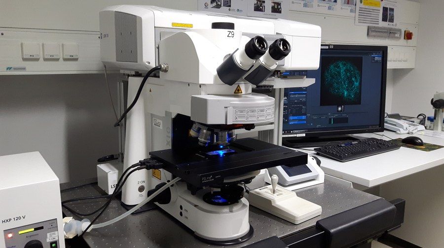
- Microdissection and contact-free sample collection (PALM MicroBeam LCM by ZEISS)
- High-resolution microscopy (Airyscan and Airyscan2 by ZEISS)
- Whole slide scanner as a service (AxioScanZ.1 by ZEISS)
- Optical sectioning by structured illumination (ApoTome2 by ZEISS)
- Cleared-sample imaging
In addition sophisticated image processing can be accomplished on high-end graphics machines using specialized software packages (ZEISS arivis Pro, ZEISS ZEN, Fiji, SVI Huygens etc.).
Our services include :
-
Support and user training for state-of-the-art microscopy techniques
-
Support of advanced microscopy techniques e.g. FLIM, FRAP, FRET, FCS, TIRF, 2P and laser microdissection.
-
Providing access to high-end acquisition and analysis workstations.
-
Instrument and workstation maintenance to ensure quality and efficiency.
Full Service Slide Scanning

In addition the facility offers full service on an automated slide scanner microscope, the Zeiss Axio Scan.Z1. Our expert staff will run the scans to digitize your specimens for reproducible data. The system features automated image acquisition of multichannel fluorescent as well as brightfield slides. See here for more information.