Dec 17, 2020
High resolution 3D microscopy enables to reconstruct the highways of insulin transport

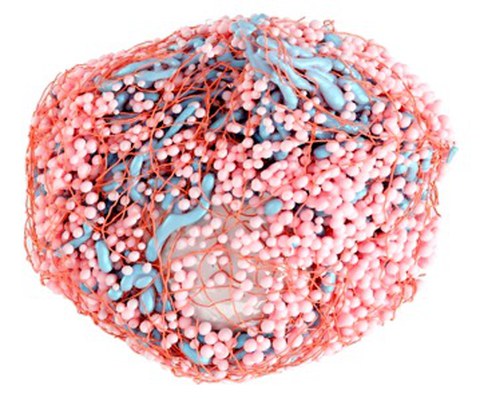
3D rendering (created by Deborah Schmidt) of one beta cell with insulin secretory granules (light orange), mitochondria (blue), nucleus (white) and microtubules (dark orange).
Microtubules are a part of cytoskeleton and act as highways for the transport of vesicles within the cell. However, their role in insulin transport and secretion is currently under debate. Now, an international team led by scientists of the Paul-Langerhans-Institute Dresden, a partner of the German Center of Diabetes Research (DZD), together with the CSBD, MPI-CBG, Janelia Research Campus, CMCB, Fondazione Human Technopole as well as the EPFL used high resolution 3D electron microscopy to image insulin secreting pancreatic islet beta cells in their entirety at an unprecedented resolution and reconstructed all insulin secretory granules, microtubules, mitochondria, Golgi apparati, and centrioles to generate a comprehensive spatial map of microtubule-organelle interactions. The outcome of this highly collaborative project was now published in the renowned journal “Journal of Cell Biology”.
Insulin is the major blood glucose lowering hormone of our body. It is stored within tiny vesicles called insulin secretory granules in the beta cells of the endocrine pancreas. Each beta cell contains several thousand insulin secretory granules. When blood glucose levels rise after a meal these insulin secretory granules fuse with the plasma membrane of the beta cell allowing the release of insulin in the blood stream. Deficient insulin secretion causes diabetes, the most common metabolic disease affecting hundreds of millions of people worldwide.
All cells, including beta cells, contain a so-called cytoskeleton. Part of this cytoskeleton are microtubules, thin filaments that build the internal scaffold of mammalian cells and can act as the tracks for the intracellular transport of vesicles. In beta cells, in particular, the microtubules are involved in the transport of the insulin secretory granules toward the plasma membrane in order to secrete insulin. However, there is also data indicating that microtubules might hinder insulin transport, especially when blood glucose levels are low. To date there has not been a full 3D reconstruction of the microtubule network in beta cells and thus lack of detailed information about its interaction with insulin secretory granules. These data, on the other hand, would provide key insight into how microtubules help regulate insulin transport and secretion.
“Microtubules are tiny filaments with a diameter of 25 nm, whereas beta cells can be up to a thousand times bigger. Combining high resolution to resolve microtubules with large volume imaging of whole beta cells is a great challenge and could not have been achieved by light microscopy methods. We therefore decided to collaborate with the Hess lab (Janelia Research Campus, USA), which has developed an enhanced 3D electron microscopy method (Focused Ion Beam Scanning Electron Microscopy (FIB-SEM) enabling the stable combination of high resolution and large volume imaging” explains Dr. Andreas Müller, the postdoc in the Solimena lab of the Paul-Langerhans-Institute Dresden and first author of the study. “Our pancreatic islet samples were continuously imaged 24 hours/day in this microscope for 4 weeks and the huge amount of collected data was used for computer-annotation and digital 3D-rendering. This remarkable feat eventually enabled us to obtain the first three-dimensional reconstructions of complete microtubule networks in primary mouse beta cells, and in mammalian cells in general, together with evidence regarding their importance for insulin secretory granule positioning and thus their supportive role in insulin secretion”.
In total, the researchers managed to reconstruct all microtubules as well as beta cell intracellular organelles such as the insulin SGs, mitochondria, centrioles with axonemes of the primary cilia, nuclei, the Golgi apparatus and plasma membrane of 7 complete beta cells (3 for the low glucose and 4 for the high glucose condition) “In most cell-types microtubules irradiate from small structures called centrioles located in the middle of the cells forming a radial network comparable to that of a bike wheel. However, the microtubule network of beta cell is different, as very few microtubules are either connected to the centrioles or to the Golgi apparatus, from which microtubules may also originate, and are instead freely positioned within the cytosol in what, at first glance, appears as a chaotic network”, summarizes Andreas Müller. “However, detailed computation of the 3D images surprisingly showed, that microtubules are enriched together with insulin secretory granules close to the plasma membrane of the beta cell. Actually, we found that many insulin granules are directly connected with microtubules, thus indicating that the latter help to locate a high amount of insulin near the plasma membrane, even when the beta cell is not stimulated by elevated blood glucose levels.”
“Another special feature of our study is the data accessibility,” says Deborah Schmidt, computational scientist at the Center for Systems Biology Dresden and second author on the paper. “We developed a plugin for the open source software FIJI so that users can overlay raw image data and organelle annotations and even generate graphs showing interactions among different compartments within cells.” “Furthermore”, adds Andreas Müller, “one large volume containing several beta cells is available via the open access platform “openorganelle”, thus enabling scientists to browse through further elaborate the data and share the results with colleagues to gain insights into the inner structures and function of beta cells besides the microtubules.”
Links
Segmentation masks and crops of analyzed beta cells:
https://cloud.mpi-cbg.de/index.php/s/UJopHTRuh6f4wR8.
FIJI plugin BetaSeg Viewer:
https://sites.imagej.net/betaseg
Full resolution beta cell volume for browsing:
https://openorganelle.janelia.org/
Original Publication:
Müller A, Schmidt D, Shan Xu C, Pang S, Verner D’Costa J, Kretschmar S, Münster C, Kurth T, Jug F, Weigert M, Hess HF, Solimena M., Three-dimensional Fib-Sem reconstruction of microtubule-organelle interaction in whole primary mouse beta cells
https://doi.org/10.1083/jcb.202010039
Journal of Cell Biology Article
