Forschung AG Schubert

In vitro Krankheitsmodellierung in mikrofluidischen Systemen
In Kollaboration mit dem Fraunhofer IWS (Dr. F. Sonntag)
https://www.iws.fraunhofer.de/de/technologiefelder/additive-fertigung-und-oberflaechentechnik/mikro-biosystemtechnik.html)
Kontakt: Dr. Mario Schubert, Prof. Dr. Kaomei Guan
Mitarbeiter: Dr. Mario Schubert, M.Sc. Oliver Gamm, M.Sc. Yuliya Dzekhtsiarova
In diesem Projekt haben wir in Kollaboration mit dem Fraunhofer IWS mikrophysiologische Zellkulturchips für die Kultivierung von aus humanen induzierten pluripotenten Stammzellen differenzierten Kardiomyozyten (iPS-Kardiomyozyten) etabliert. Die Zellkulturchips bilden dabei ein geschlossenes System, in dem das Kulturmedium durch integrierte Mikropumpen zirkuliert. Eine komplexe Steuerung ermöglicht nicht nur die Regulierung der Flussraten, sondern ebenso der Sauerstofflevel in der Kulturkammer der iPS-Kardiomyozyten. Während der Kultivierung können die iPS-Kardiomyozyten mittels Durchlicht- sowie Fluoreszenzmikroskopie dokumentiert und so beispielsweise mittels High-Speed-Imaging die kontraktile Funktion charakterisiert werden. So können wir komplexe biologische Vorgänge rekapitulieren und pathophysiologische Szenarien in vitro nachbilden. Unser aktueller Fokus liegt in der Simulation eines akuten Myokardinfarktes und der damit verbundenen Hypoxie-Reperfusionsschädigung der Kardiomyozyten.
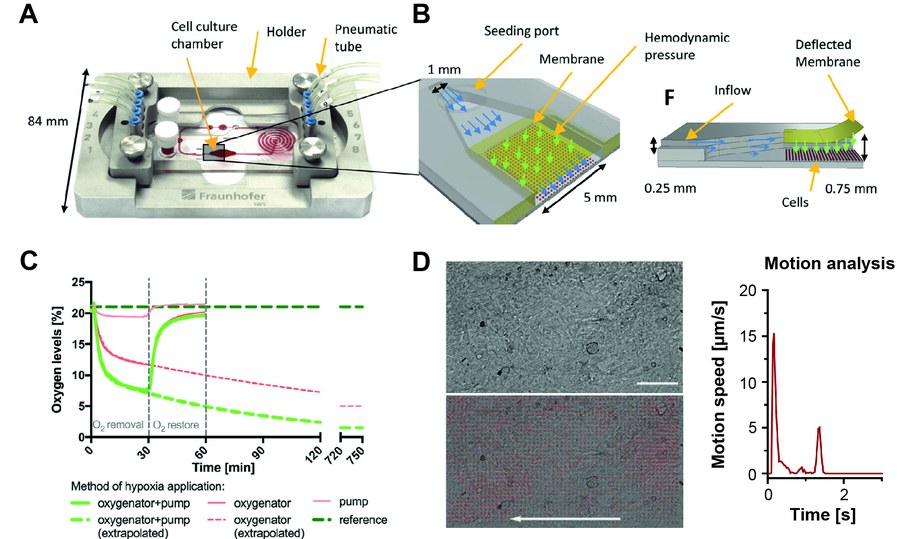
Abbildung 1: Mikrophysiologisches System (MPS) zur Kultivierung von iPS-Kardiomyozyten und zur Modellierung pathologischer Zustände. A, B, Aufbau des MPS Chips in Support (A) und Detaildarstellung der Zellkulturkammer (B) mit semipermeabler Membran, Mediumfluss und Induzierung hämodynamischer Stimulation. C, Regulation des Sauerstoffgehalts im Kulturmedium zur Modellierung hypoxischer Zustände. D, Video-basierte Analyse der Kardiomyozytenfunktion im MPS anhand der Kontraktionsbewegungen (Kolanowski et al., 2020).
Etablierung eines humanen Multi-Organ-on-a-Chip Modells zur Untersuchung der Monozyten- und Makrophagenfunktion nach Ischämie-Reperfusionsschädigung
Das Projekt wird vom Bundesinstitut für Risikobewertung unter dem Förderkennzeichen 60-0102-01#00067 - P639 gefördert.
Kontakt: Dr. Mario Schubert, Prof. Dr. Kaomei Guan
Mitarbeiter: M.Sc. Shakthi Arun, M.Sc. Oliver Gamm, B.Sc. Pranotee Gawade, Paul Beck
Kooperationen:
Prof. Dr. Andreas Richter (Chair of Microsystems, TU Dresden)
Dr. Mathias Busek (Hybrid Technology Hub, Universität Oslo)
Der akute Myokardinfarkt (MI) und die resultierende Entwicklung von Herzversagen (HF) ist eine der häufigsten Ursachen für Einschränkung der Lebensqualität und Tod, und stellt eine große medizinische Herausforderung dar. Die Reaktion des Immunsystems, insbesondere der Monozyten und Makrophagen, ist von kritischer Bedeutung für das Ausmaß sowie die Reparatur der Gewebeschädigung und stellt einen wichtigen Ansatzpunkt zur Entwicklung neuer Therapien dar. Tiermodelle können die Immunreaktion nach MI nur bedingt abbilden und gehen mit einer substantiellen Zustandsverschlechterung und Tod der Tiere einher. Mit der Entwicklung eines Multi-Organ-on-a-Chip-Systems (MOoC) auf Basis humaner Zellmodelle wollen wir diese Lücke in der translationalen Forschung schließen und Tiermodelle ersetzen. Das MOoC-System ermöglicht die Kultivierung eines komplexen Herzgewebemodells; bestehend aus Kardiomyozyten, Fibroblasten sowie residenten Makrophagen; die Integration einer Endothelbarriere und die Zirkulation von Monozyten und Makrophagen. Die durch den MI ausgelöste Ischämie-Reperfusionsschädigung (I/R) soll mit dem MOoC simuliert und die Infiltration von Monozyten und Makrophagen in das Herzgewebe untersucht werden. So kann die Rolle verschiedener Monozyten- und Makrophagen-Subtypen bei der Entzündungsreaktion, Gewebereparatur und Entstehung von HF analysiert werden. Durch den Einsatz humaner induzierter pluripotenter Stammzellen zur Herstellung der eingesetzten Zelltypen basiert das MOoC komplett auf humanen Zellmodellen. Das MOoC-System zur in vitro-Simulation der Monozyten- und Makrophageninfiltration nach I/R bietet, über die Fragestellung der Herzschädigung nach MI hinaus, großes Potential zum Ersatz von Tiermodellen in verschiedenen Forschungsbereichen.
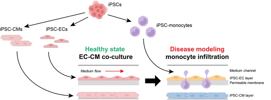
Abbildung 2: Humanes Multi-Organ-on-Chip-System (MOoC) zur Untersuchung von Monozyteninfiltration in geschädigtes Herzgewebe basierend auf induzierten pluripotenten Stammzellen (iPSCs). Kardiomyozyten (iPSC-CM), Endothelzellen (iPSC-EC) und Monozyten werden aus iPSCs differenziert und in einem MOoC System eingesetzt, wobei mittels EC eine funktionelle Membranbarriere und mittels CMs Herzmuskelgewebe dargestellt wird. Durch die Inkubation unter Hypoxie werden EC und CMs geschädigt. Durch die Applikation von iPSC-Monozyten in das im Chip zirkulierende Medium wollen wir deren Infiltration in das Herzgewebe, wie nach einem Myokardinfarkt, nachstellen und untersuchen.
Interaktion von Kardiomyozyten und kardialer Fibroblasten in Entwicklung und Pathophysiologie
Ehemals gefördert durch MeDDrive Grant der TU Dresden (zu Dr. M. Schubert).
Kontakt: Dr. Mario Schubert, Prof. Dr. Kaomei Guan
Mitarbeiter: Wilhelm Wenke, Paul Josef Conrad Beck
Kardiale Fibroblasten nehmen eine maßgebliche Rolle für physiologische und pathophysiologische Prozesse im Herzen ein, beispielsweise im Rahmen der Herzentwicklung oder der Ausbildung kardialer Fibrose. Die zugrundeliegenden molekularen Mechanismen der Zell-Zell-Interaktion zwischen verschiedenen Zelltypen des Herzens sind bislang nur unzureichend erforscht. Eine bessere Kenntnis auf diesem Gebiet könnte jedoch sehr wahrscheinlich neue Ansätze zur Entwicklung neuer Therapien und Identifizierung neuer pharmakologischer Zielstrukturen liefern. Aus diesem Grund beschäftigen wir uns mit der Wechselwirkung humaner iPS-Kardiomyozyten und kardialer Fibroblasten. In unseren Studien kommen sowohl 2D-Zellkulturmodelle als auch 3D-Gewebesysteme, Engineered Heat Tissues oder kardiale Organoide, mit mehreren kardialen Zelltypen (Kardiomyozyten, Fibroblasten, Endothelzellen) zum Einsatz. Die Herstellung von Engineered Heart Tissues als Herzgewebemodell aus iPS-Kardiomyozyten und humanen ventrikulären Fibroblasten ermöglich es uns beispielsweise, die Entstehung von Fibrose und deren Auswirkung auf die Gewebefunktion zu untersuchen.
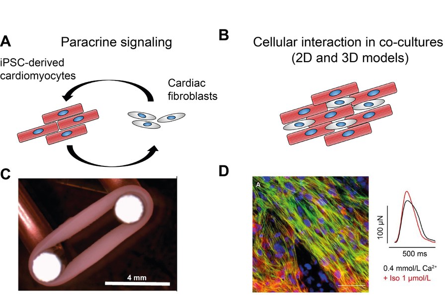
Abbildung 3: Untersuchung der Kardiomyozyten-Fibroblasten Interaktion. A, B, Der bidirektionale Crosstalk beider Zelltypen finden durch parakrine Signalwege (A), durch die Sezernierung von löslicher Faktoren oder extrazellulärer Matrix, oder durch direkte Zell-Zell-Kontakte (B) statt. C, D, Diese Zell-Zell-Interaktionen untersuchen wir beispielsweise in Engineered Heart Tissues, kleine Muskelringe, die mittels Kollagenmatrices geformt und auf Stretchern kultiviert werden (C). Diese Gewebe ermöglichen sowohl molekulare Analysen der Protein- und Genexpression, als auch die Messungen der Kontraktionskraft unter pharmakologischer Stimulation (D).
Maturierung von iPS-Kardiomyozyten in 2D und 3D Modellen für Wirkstofftestung und Krankheitsmodellierung
Ehemals gefördert durch MeDDrive Start der TU Dresden (zu Dr. M. Schubert)
Kontakt: Dr. Mario Schubert, Prof. Dr. Kaomei Guan
Mitarbeiter: Paul Josef Conrad Beck, M.Sc. Oliver Gamm, M.Sc. Yuliya Dzekhtsiarova, B.Sc. Isha Bansal
Eine wesentliche Limitierung bei der Entwicklung neuer Wirkstoffe stellt die mangelnde Translationsfähigkeit prä-klinischer Therapiekonzepte aus Mausmodellen hin zur klinischen Anwendung im Patienten dar. Die Etablierung humaner Modellsysteme ist daher ein Schlüsselbaustein für zukünftige Arzneimittelentwicklung und zur Reduktion von Tierversuchen. Die Herstellung humaner Kardiomyozyten durch kardiale Differenzierung von induzierten pluripotenten Stammzellen (iPS-Kardiomyozyten) liefert große Mengen humaner Herzmuskelzellen als Modellsysteme für biomedizinische Forschung und Wirkstoffscreenings. Eine Limitation im Hinblick auf das translationale Potential stellt jedoch der embryonale, unreife Phänotyp der iPS-Kardiomyozyten dar welcher die Risikoevaluation kardiotoxischer oder arrhythmogener Wirkstoffe sowie die Etablierung von in vitro Krankheitsmodellen erschwert. Der Fokus in diesem Projekt liegt darauf, die iPS-Kardiomyozyten im Hinblick auf deren Funktionalität, Stoffwechsel und Zellstruktur so zu reifen, dass sie den Eigenschaften adulter Herzmuskelzellen rekapitulieren. Dazu werden iPS-Kardiomyozyten in 2D- und 3D-Zellmodellen in Gegenwart verschiedener Stimuli kultiviert und die Zellen funktionell, metabolisch und strukturell charakterisiert.
Methoden:
-
Differenzierung humaner Kardiomyozyten, Monocyten, Makrophagen und Endothelzellen aus induzierten pluripotenten Stammzellen
-
Genom-Editierung mittels CRISPR-Cas9 Technologie
-
Expressionsanalysen (Real-Time PCR, Western Blot, Immunfärbungen)
-
Fluoreszenzmikroskopie
-
Durchflusszytometrie
-
Analyse des Zellmetabolismus: Seahorse Assays, Oroboros
-
Video-basierte Kontraktionsanalyse
-
Tissue Engineering: Engineered Heart Tissues
-
Lebendzell-Fluoreszenzmikroskopie (Calciumtransienten, Calcium-Sparks, Aktionspotentiale)
-
Mikrophysiologische Systeme
Mitarbeiter:innen
Dr. Mario Schubert – Arbeitsgruppenleiter
Telefon: +49 351 458 6421
Email:
Paul Beck – MD Student
Philipp Hartmann – MD Student
Oliver Gamm – PhD Student
Shakthi Arun - PhD Studentin
Isha Bansal - Masterstudentin
Pranotee Gawade - Masterstudentin
Niclas Speri - MD Student
Wilhelm Wenke – MD Student
Sinah Hansen – MD Studentin
Alumni:
Robert-Patrick Steiner – MD Thesis
Marcel Hasse - MD Thesis