Cell culture
Cells interact with each other and with diverse biomaterials. The research of cells and their behavior are therefore of great interest, both in cell culture (in vitro), as well as in animal models (in vivo).
Cell Culture
We use cells from different sources for our research projects. As S1-certified laboratory we work with cell lines, but also with primary cells isolated directly from human and animal sources. In addition to classical cell culture we have established a variety of co-culture models. In new studies on "Green bioprinting" we also use cells of plant origin.
- hTERT MSC, human mesenchymal cell line
- SAOS-2, human osteosarcoma cell line
- C2C12, mouse myoblast cell line
- ST-2, mouse bone marrow stromal cell line
- L929, mouse fibroblast cell line
- RAW 264.7, mouse macrophage cell line
- more mouse and rat cell lines
- MSC from bone marrow
- MSC from adipose tissue
- MSC from reaming debris
- MSC from tendon
- human chondrocytes
- human osteoblasts from femoral heads
- human dermal microvascular endothelial cells (HDMEC)
- human umbilical vein endothelial cells (HUVEC)
- human dermal fibroblasts
- human peripheral blood mononuclear cells (PBMC)
- human dental pulp stem cells
•bovine MSC, isolated from bone marrow
•MSC from bone marrow of rabbits and rats
•porcine MSC, isolated from bone marrow
- basilicium cells
- algae
Special Cell Culture Conditions
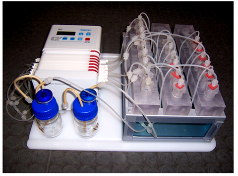
We are equipped with cell culture incubators and sterile culture hoods that allow a strict separation of mycoplasm-free cultures and fresh primary cells. All incubators maintain the standard concentration of 5 % CO2, and three incubators also allow culturing under hypoxic conditions. Numerous perfusion systems, from commercially available to custom-made modules, enable the dynamic cultivation of cells in 2D and 3D scaffolds.
Analytical Methods
The characterization of cells can be carried out using various methods of analysis. To analyze cells, we apply methods such as enzyme linked immunosorbent assay (ELISA) for collagen II, BMP-2, VEGF and other proteins. Furthermore, we routinely use colorimetric and fluorescence spectrometric assays to quantify the DNA content and enzyme activities (LDH, ALP, TRAP, GPDH, CAII and more) in cell lysates. The expression of mRNA is analyzed using RT-PCR (qPCR and semi-quantitative PCR). A variety of other techniques such as histology or different immune fluorescence stainings are also established. If you are interested in a special process or its implementation, please do not hesitate to contact us.


