Liquid ventilation and OCT
Up to now, the quality of three-dimensional OCT images of lung tissue is mainly affected by two effects. Firstly, there is a limited penetration depth of near-infrared light into tissue, secondly, alveolar structures are displayed incorrectly with flattened wall surfaces parallel to the beam direction due to total reflection at the tissue-air interface. Due to the refractive index differences of tissue and air-filled alveoli, scattering losses are high and image quality is decreased. Both effects can be significantly reduced by using an appropriate liquid, as the refractive index inside the alveoli can be matched to the one of the surrounding tissue, whereby scattering losses are strongly reduced. Previous studies of our research group have shown these positive effects for the imaging of isolated and fixed rabbit lungs.
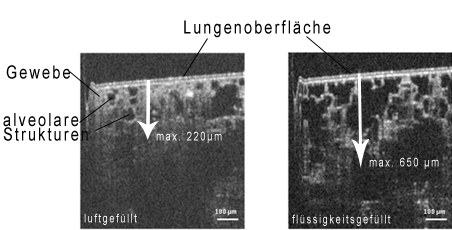
Comparison of two OCT cross sections with different breathing media. Left: air-filled lung, the penetration depth for imaging is limited to about 0.2 mm, the alveolar structures are partly hard to recognize. Right: Liquid-filled lung, the penetration depth is almost three times greater and the tissue structure is clearly visible.
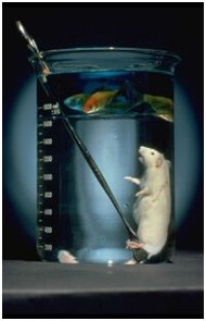
Experiment by Golland and Clark in 1966; a mouse is fixed in a perfluorocarbon. On top, there is a layer of water with fish. There is no mixing due to the different densities and the chemical inertness of perfluorocarbon. The mouse ventilates only PFC, absorbs the bounded oxygen and emits carbon dioxide into the liquid.
The model of fixated rabbit lung is not sufficient to study dynamic changes of lung tissue during ventilation and to establish detailed numeric models. Therefore, we develop a liquid ventilation device, which allows transferring the advantages for imaging to an in vivo measuring situation. As breathing medium perfluorocarbons are used, which transport the vital breathing gases oxygen and carbon dioxide into and out of the lung. The concept of liquid ventilation is already known for 50 years and has been constantly enhanced; however, it has never been widely established in clinical routine due to the high technical effort and the mostly unexplored interactions of the liquid breathing media and the human organism. Nevertheless, liquid ventilation is used in some clinics as special therapy in neonatology. Besides the enhanced imaging for OCT, this project shall deal with further medical questions of liquid ventilation, such as drug carriers or inflammation inhibition.
Funding
German Research Foundation (DFG - KO 1814/6-1 und 6-2)
Kooperationen
Prof. Dr. Wolfgang Kübler, St. Michael's Hospital Toronto
Dr. Hannah Nickles, Institute of Physiology, Charité - Universitätsmedizin Berlin
Contact
 © Julia Walther, KSM
Mr Dr. rer. medic. Christian Schnabel
Send encrypted email via the SecureMail portal (for TUD external users only).
Clinical Sensoring and Monitoring
Clinical Sensoring and Monitoring
Visiting address:
Medizinisch-Theoretisches Zentrum (MTZ - Haus 91) Fiedlerstraße 42
01307 Dresden