Aug 15, 2024
Thalamic Brain Dysfunction in Dyslexia: A Breakthrough in Understanding the Most Common Learning Disorder
A research team headed by neuroscientist Prof. Katharina von Kriegstein at TUD Dresden University of Technology confirms that developmental dyslexia is linked to changes in the function and structure of a specific part of the human brain known as the visual thalamus. Their findings belong to the largest and most comprehensive high-resolution MRI study targeting this area. The study has now been published in the journal ‘Brain’.
The visual thalamus is a key region connecting the eyes with the cerebral cortex. It consists of two distinct subdivisions with distinct roles in visual information processing: one smaller part specialized in detecting motion and rapidly changing visual information, and one larger part focused on color processing. Earlier post-mortem studies suggested alterations in dyslexia specifically within the smaller motion-sensitive subdivision of the visual thalamus. However, these findings were never confirmed in living humans, and the contribution of these alterations to dyslexia symptoms remained uncertain. This is because the subdivisions of the visual thalamus are tiny, with the smaller one only about the size of a peppercorn, and because they are located deep in the brain. This makes it exceptionally difficult to assess these structures with conventional magnetic resonance imaging (MRI). To overcome this challenge, for the current study, the researchers conducted a series of novel experiments on a specialized MRI system at the Max Planck Institute of Human Cognitive and Brain Sciences (MPI-CBS) in Leipzig. Equipped with a powerful magnet, this MRI system made it possible to investigate the visual thalamus at an unprecedented level of detail in living humans. To maximize the number of participants with dyslexia who met the stringent safety standards for these MRI examinations, the authors were supported by the Bundesverband für Legasthenie und Dyskalkulie e.V. (English: Federal Association for Dyslexia and Dyscalculia; https://www.bvl-legasthenie.de/, short: BVL), who expanded the recruitment efforts throughout Germany. The study results, based on a sample of 25 individuals with dyslexia and 24 matched controls, revealed that dyslexia is linked to alterations in both the function and structure of the motion-sensitive subdivision of the visual thalamus. Furthermore, these alterations were associated with dyslexia symptoms, particularly among male individuals with dyslexia, suggesting potential sex-differences in the pathology of dyslexia.

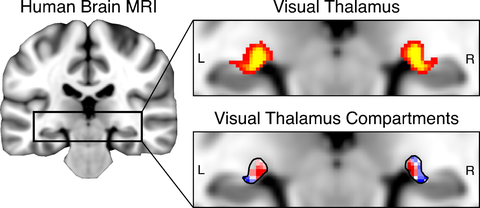
The left panel shows an overview MRI of the human brain. The upper panel insert shows the location of the visual thalamus. Yellow shading indicates a higher number of participants with the visual thalamus located in this position. The bottom panel illustrates the two subdivisions of the visual thalamus: the motion-sensitive subdivision, which is altered in individuals with dyslexia, is shown in red.
Dr. Christa Müller-Axt, Postdoctoral Researcher at the Chair of Cognitive and Clinical Neuroscience at TU Dresden explains the impact of their findings: “We confirmed a long-standing hypothesis on brain dysfunction in developmental dyslexia, revealing alterations in the visual thalamus – a tiny structure deep inside the brain. For decades, investigating this structure and its subdivisions in living humans was impossible. However, we now present the largest and most comprehensive high-resolution MRI study targeting this area. Partnering with the BVL has been instrumental in achieving this pivotal advance in our understanding of the disorder, which could profoundly impact future diagnostic and treatment strategies.”
With a prevalence of 5-10%, developmental dyslexia is the most common learning disorder, affecting millions of people worldwide. Individuals with dyslexia face severe difficulties in developing adequate literacy skills, often accompanied by ongoing struggles in school, limitations in the workforce, and emotional difficulties. Despite the high prevalence, the neurobiological causes of dyslexia remain elusive. Partly due to the limited availability and increased expense of specialized MRI systems, most neuroscience research has focused on the cerebral cortex in explaining dyslexia. Consequently, the role of the thalamus in dyslexia has remained largely unexplored. Moreover, early post-mortem studies on thalamic alterations in dyslexia were limited to a few patients, leaving uncertainty about whether these alterations are common to all individuals with dyslexia or only to a subset. “Our discovery of thalamic alterations in a large sample of individuals with dyslexia suggests that these changes may be more prevalent in the disorder's pathology than previously recognized. This finding paves the way for further research aimed at gaining a more comprehensive understanding of the brain mechanisms underlying dyslexia. Importantly, our results also show that thalamic alterations are associated with reading-related difficulties, specifically in male individuals with dyslexia. This underscores the need to explore how thalamic changes uniquely impact behavior, pivotal for effectively developing therapy and intervention strategies for dyslexia”, concludes Prof. Katharina von Kriegstein, Chair of Cognitive and Clinical Neuroscience at TU Dresden.
Original publication:
Christa Müller-Axt, Louise Kauffmann, Cornelius Eichner and Katharina von Kriegstein. Dysfunction of the magnocellular subdivision of the visual thalamus in developmental dyslexia. Brain. https://doi.org/10.1093/brain/awae235
Contact:
Prof. Katharina von Kriegstein
Chair of Cognitive and Clinical Neuroscience
TUD Dresden University of Technology
Email:
Tel: +49 351 463-43901
Dr. Christa Müller-Axt
Postdoctoral Researcher
Chair of Cognitive and Clinical Neuroscience
TUD Dresden University of Technology
Email:

