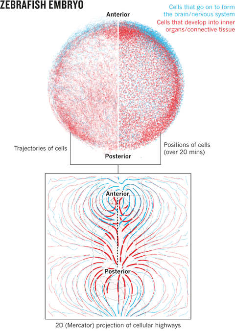
Analysis of cell migration patterns in the zebrafish embryo
The zebrafish danio rerio is one of the most commonly used model organism, especially in the field of developmental biology. During gastrulation (period in embryonic development) the first, very complex deformations of the embryonic tissue take place, at the same time cells (blastomeres) differentiate into three different germ lines which later form all the organs and tissue of the fish. We study the processes of tissue reorganisation (emboly, cell sorting, thinning etc.) through different types of cell movements like involution, epiboly, convergence&extension, on the basis of light sheet microscopy. There, for the first time in toto imaging of the developing embryos in time and space has been made available, with close to optimal conditions for the studied specimen. Our project involves raw data processing, visualisation and statistical analysis of the cellular processes we achieve from the multidimensional time-lapse image data
Involved scientists
- Konstantin Thierbach
- Ingo Röder
Publications
Collaboration partners
- Jan Huisken (Max Planck Institute of Molecular Cell Biology and Genetics, Pfotenhauerstr. 108, 01307 Dresden, Germany and Morgridge Institute for Research, Madison, Wisconsin 53715, United States of America)
- Gopi Shah (Max Planck Institute of Molecular Cell Biology and Genetics, Pfotenhauerstr. 108, 01307 Dresden, Germany and Cancer Research UK Cambridge Institute, University of Cambridge, Robinson way, CB20RE Cambridge, UK)
- Nico Scherf (Max Planck Institute of Molecular Cell Biology and Genetics, Pfotenhauerstr. 108, 01307 Dresden, Germany and Max Planck Institute for Human Cognitive and Brain Sciences, Stephanstraße 1a, 04103 Leipzig, Germany)

